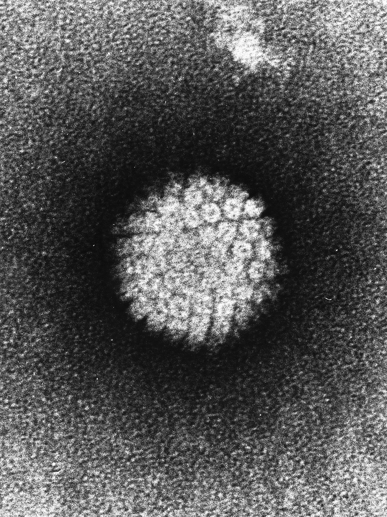
CONTACT

WOV

  • COVID-19
    • C’RONA COMIX
    • TRIBAL C’RONA COMIX
    • C’RONA COMIX II
    • C’RONA PANDEMIC COMICS
  • COMICS & Apps
    • C’RONA PANDEMIC COMICS
    • CARNIVAL OF CONTAGION
    • Confined!
    • Phantom Planet
    • The Never-Ending Battle
    • The Frozen Horror
    • Curse of the Tree-man
    • Occupied by microbes!
    • Microbe Stickers
  • Books & Audio
    • C’RONA PANDEMIC COMICS
    • PLANET OF VIRUSES
    • World of Viruses
    • Watch Your Mouth!
    • Microbe Stickers
    • CARNIVAL OF CONTAGION
    • Audio Stories
  • Microbe Images
    • Inside Viruses
    • Microbe Scientists
    • Microbe SEMS
    • Historical Images
    • Microbe Illustrations
  • Activities
  • Omaha Sci Media
  • Worlds of Connections
  • Research & Evaluation

Image wall, Microbe SEMS

Electron micrograph of 1918 influenza virus

January 9, 2013 jbrehm2
Description

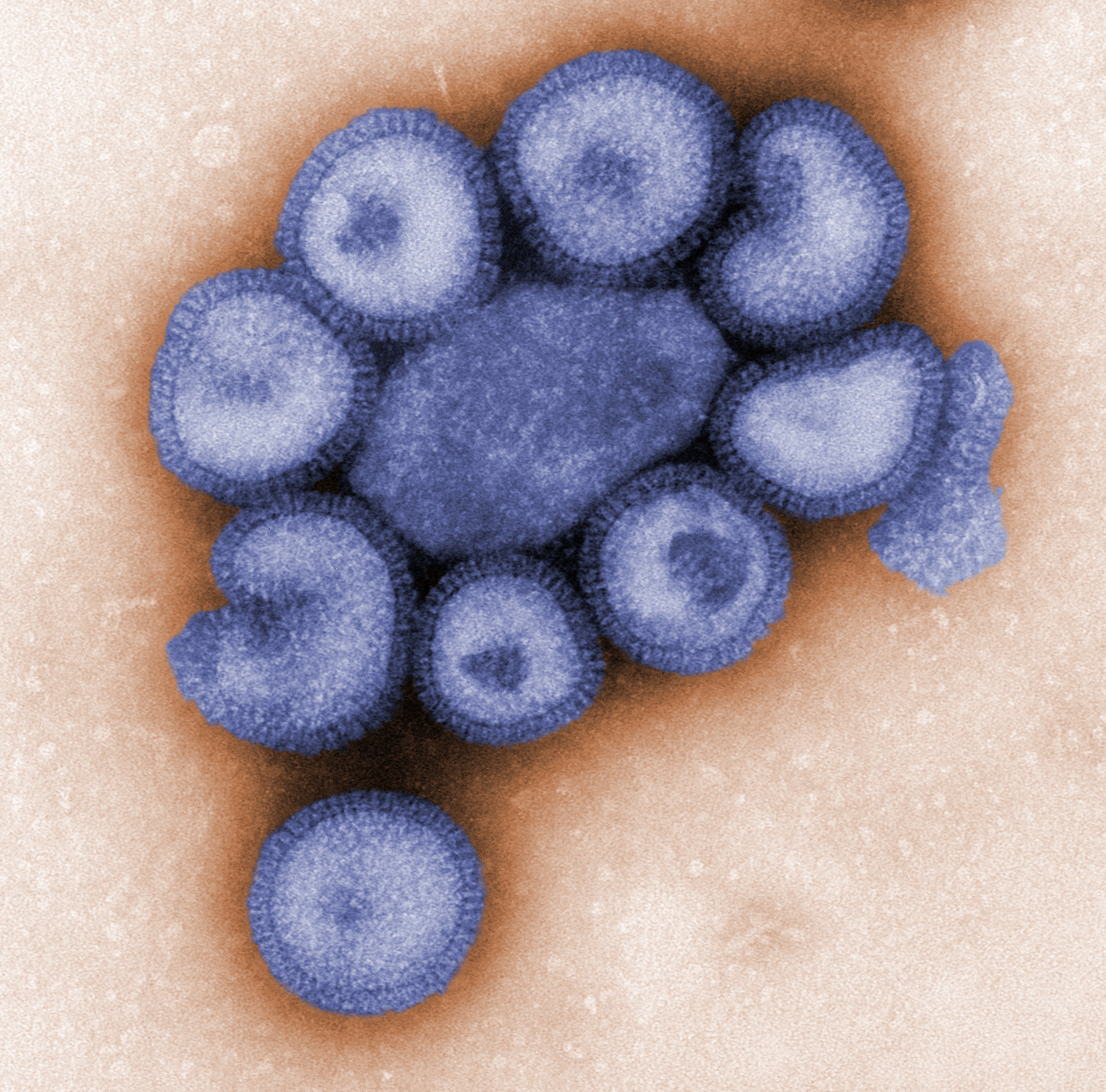
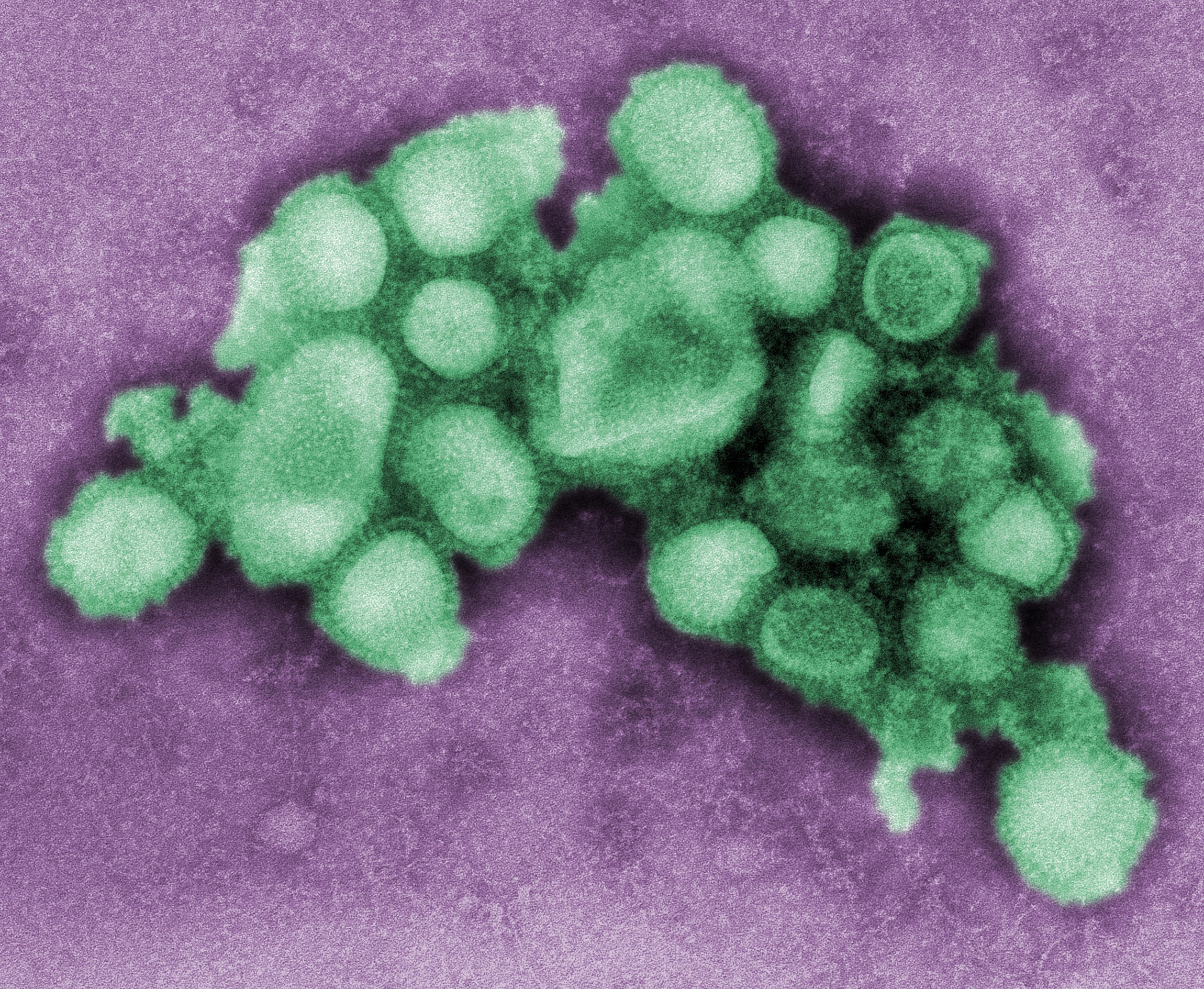
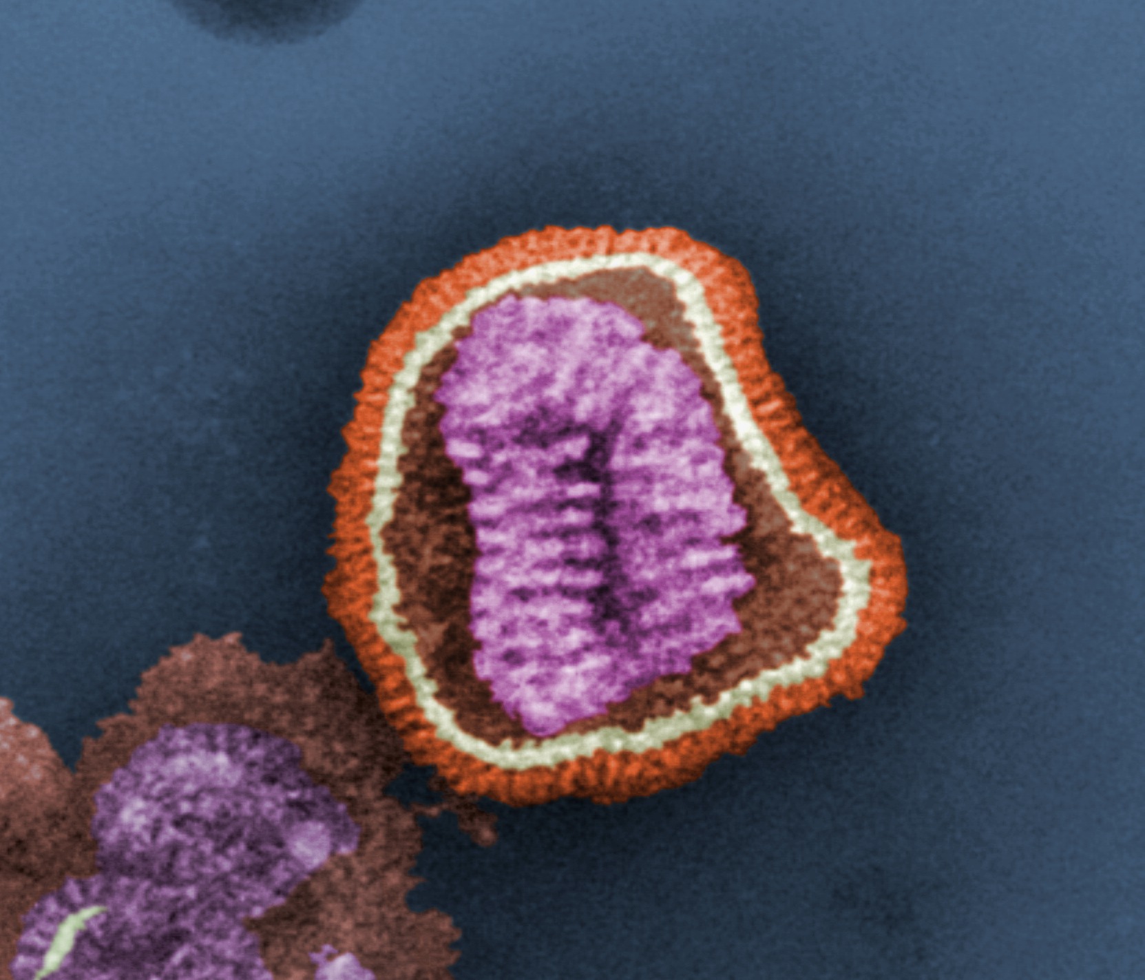
This electron micrograph shows several virus particles, or virions, of a re-created 1918 influenza virus growing in kidney cells of a dog.

Credit

Public domain/ Centers for Disease Control and Prevention/ Dr. Terrence Tumpey, content provider/ Cynthia Goldsmith, photographer/ 2005

Source

http://phil.cdc.gov/phil/imageidsearch.asp (Image ID # 11098)

Image wall, Microbe SEMS

Electron micrograph of influenza virus particles

January 9, 2013 jbrehm2
Description

Electron micrograph shows a number of influenza virus particles, or virions.

Credit

Public domain/ Centers for Disease Control and Prevention/ Dr. F. A. Murphy, content provider/ 1973

Source

http://phil.cdc.gov/phil/imageidsearch.asp (Image ID # 10072)

Image wall, Microbe SEMS

Electron micrograph of influenza virus isolated from pigs

January 9, 2013 jbrehm2
Description

This electron micrograph shows several particles, or virions, of an influenza virus that causes the disease in pigs.

Credit

Public domain/ Centers for Disease Control and Prevention/ C. S. Goldsmith and A. Balish, content providers/ 2009

Source

http://phil.cdc.gov/phil/imageidsearch.asp (Image ID # 11215)

Image wall, Microbe SEMS

Electron micrograph of influenza virus

January 9, 2013 jbrehm2
Description

This electron micrograph shows an influenza virus particle, or virion. Influenza is a single-stranded RNA virus.

Credit

Public domain/ Centers for Disease Control and Prevention/ E. L. Palmer and M. Larkin, content providers/ F. Murphy, photographer/ 1981

Source

http://phil.cdc.gov/phil/imageidsearch.asp (Image ID # 10073)

Image wall, Inside Viruses

Influenza virus (inside)

January 9, 2013 jbrehm2
Description

A view of the influenza virus in which a portion of the protein coat, or capsid, has been cut away, showing the RNA inside.

Credit

Public domain/ Centers for Disease Control and Prevention/ Douglas Jordan, content provider/ Dan Higgins, illustrator/ 2009

Source

http://phil.cdc.gov/phil/imageidsearch.asp (Image ID # 11822)

See the outside of the virus


Image wall, Inside Viruses

Influenza virus

January 9, 2013 jbrehm2
Description

A 3-D graphical representation of a generic influenza virus.

Credit

Public domain/ Centers for Disease Control and Prevention/ Douglas Jordan, content provider/ Dan Higgins, illustrator/ 2009

Source

http://phil.cdc.gov/phil/imageidsearch.asp (Image ID # 11823)

See the inside of the virus


Image wall

HPV vaccination

January 9, 2013 jbrehm2
Description

A teenage girl receives an HPV vaccination from a nurse.

Credit

Public domain/ Centers for Disease Control and Prevention, CDC/ Judy Schmidt, content provider/ James Gathany, photographer/ 2006

Source

http://phil.cdc.gov/phil/imageidsearch.asp (Image ID # 9400)

Image wall

HPV vaccination with family

January 9, 2013 jbrehm2
Description

A nurse gives an HPV vaccination to a teenage girl as her family looks on.

Credit

Public domain/ Centers for Disease Control and Prevention, CDC/ Judy Schmidt, content provider/ James Gathany, photographer/ 2006

Source

http://phil.cdc.gov/phil/imageidsearch.asp (Image ID # 9409)

Image wall, Microbe SEMS

Electron micrograph of human papillomavirus

January 9, 2013 jbrehm2
Description

This is an electron photomicrograph of human papillomavirus (HPV) occurring in human warts.

Credit

Public domain/ Laboratory of Tumor Virus Biology, National Cancer Institute/ 1986

Source

http://visualsonline.cancer.gov/details.cfm?imageid=2255

Image wall, Inside Viruses

Human papillomavirus (inside)

January 9, 2013 jbrehm2
Description

This shows the human papillomavirus (HPV), in which a portion of the protein coat, or capsid, has been cut away to reveal the DNA inside.

Credit

Permission granted for educational use/ University of Nebraska-Lincoln/ Angie Fox, illustrator/ 2010

See the outside of the virus


Posts navigation

← Previous 1 … 7 8 9 … 20 Next →

Recent Posts

  • Tribal C’rona Comix Page 5
  • Tribal C’rona Comix Page 4
  • Tribal C’rona Comix Page 3
  • Tribal C’rona Comix Page 2
  • Tribal C’rona Comix Page 1

Recent Comments

    Archives

    • December 2020
    • November 2020
    • October 2020
    • September 2020
    • August 2020
    • July 2020
    • June 2020
    • February 2015
    • December 2014
    • January 2013

    Categories

    • Apps and Activities
    • Bacterial Viruses
    • Covid-19
    • Crona Comix II
    • Curricula
    • Dengue Virus
    • Ebola Virus
    • Epidemic Simulations
    • Foot and Mouth Disease Virus
    • General
    • Hantavirus
    • Hepatitis B Virus
    • Historical Images
    • HIV
    • HPV
    • Image wall
    • Influenza Virus
    • Inside Viruses
    • Microbe Illustrations
    • Microbe Scientists
    • Microbe SEMS
    • Polio Virus
    • Rabies Virus
    • SARS Virus
    • Tribal Crona Comix
    • Uncategorized
    • West Nile Virus

    Meta

    • Log in
    • Entries feed
    • Comments feed
    • WordPress.org