University of Pittsburgh Public Health Dynamics Laboratory
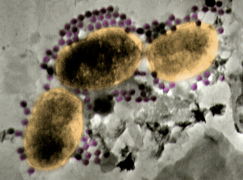
Marine viruses, shown in purple, surround potential microbes. The odds are stacked against the microbes when there are so many viruses. Willie Wilson, Bigelow Laboratory for Ocean Sciences.
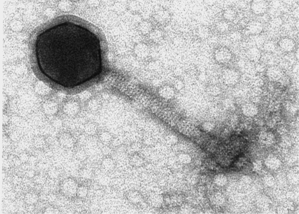
Phages are viruses that infect bacteria. They have a characteristic shape. Cyanophage photo by Willie Wilson, Bigelow Laboratory for Ocean Sciences.
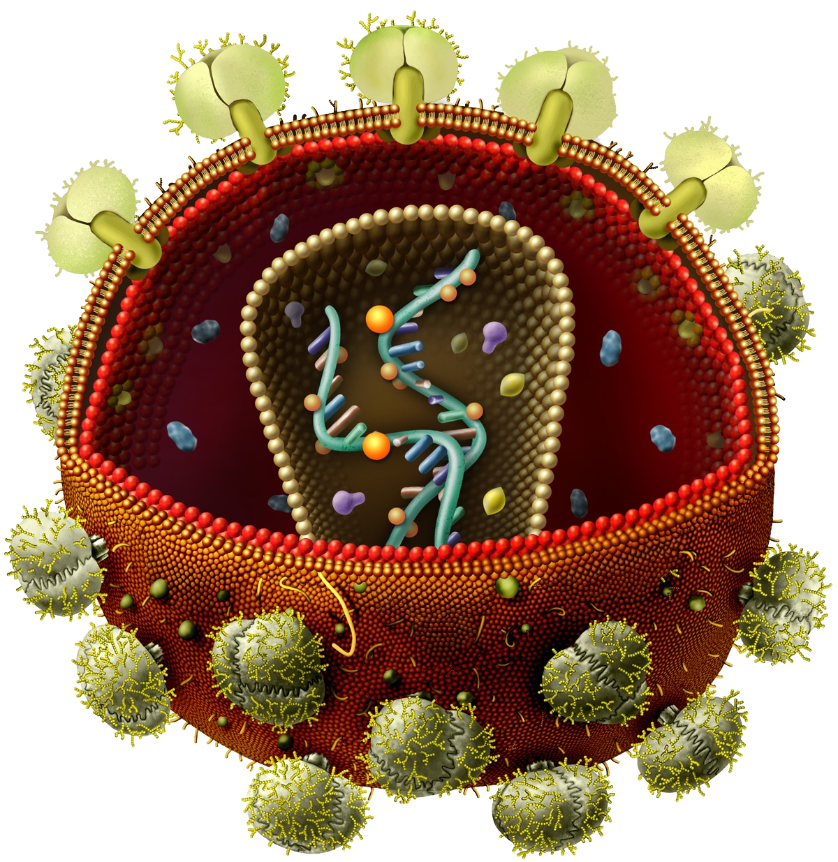
This illustration shows the interior of the human immunodeficiency virus (HIV), which causes AIDS.
Permission granted for educational use/ University of Nebraska State Museum/ Angie Fox, Illustrator/ 2010
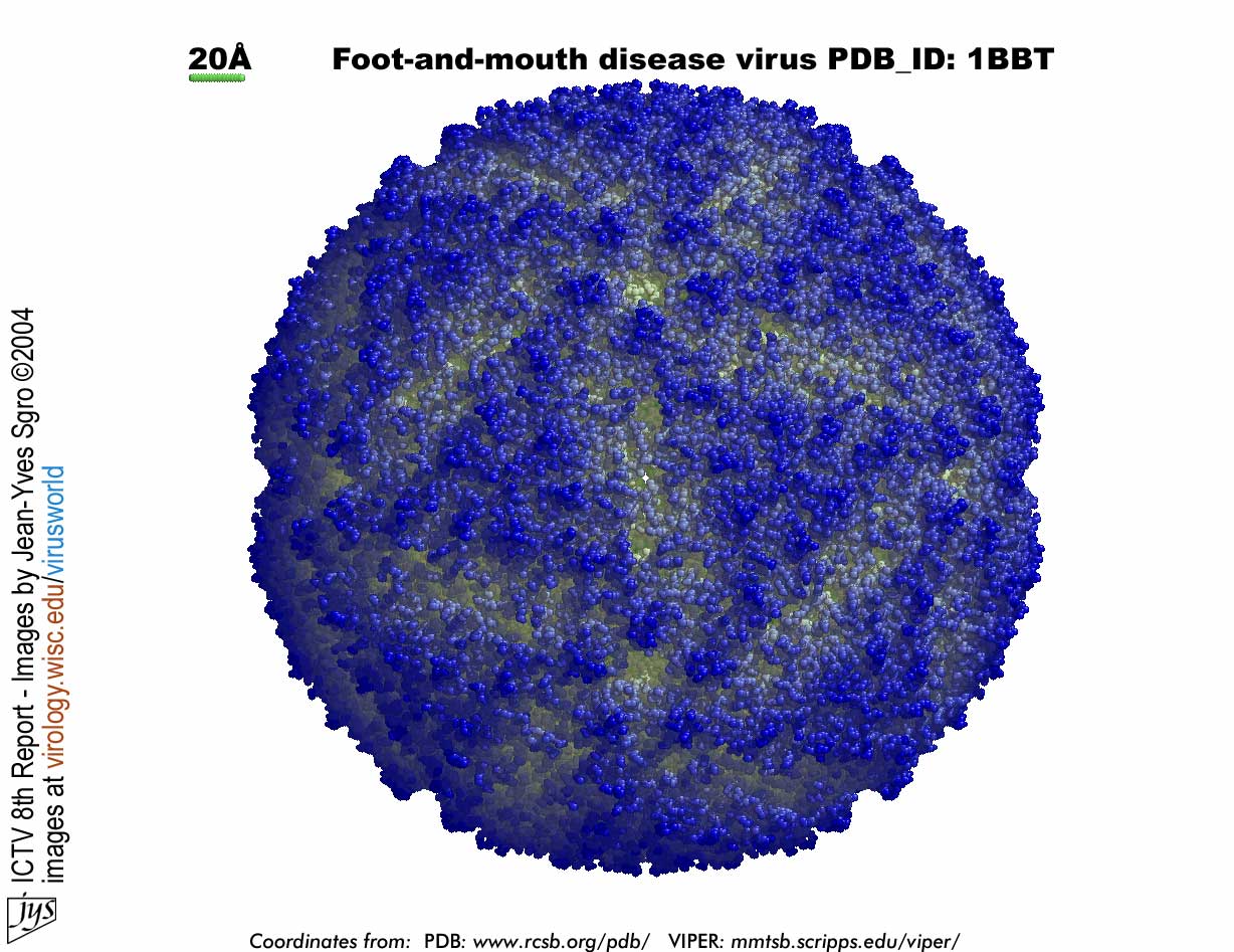
Foot-and-mouth disease viruses cause illness in cloven-hoofed animals, such as cattle, pigs, and sheep. FMD is an RNA virus.
Permission granted for educational use/ Virus World/ Institute for Molecular Virology/ University of Wisconsin-Madison/ Jean-Yves Sgro/ 2004
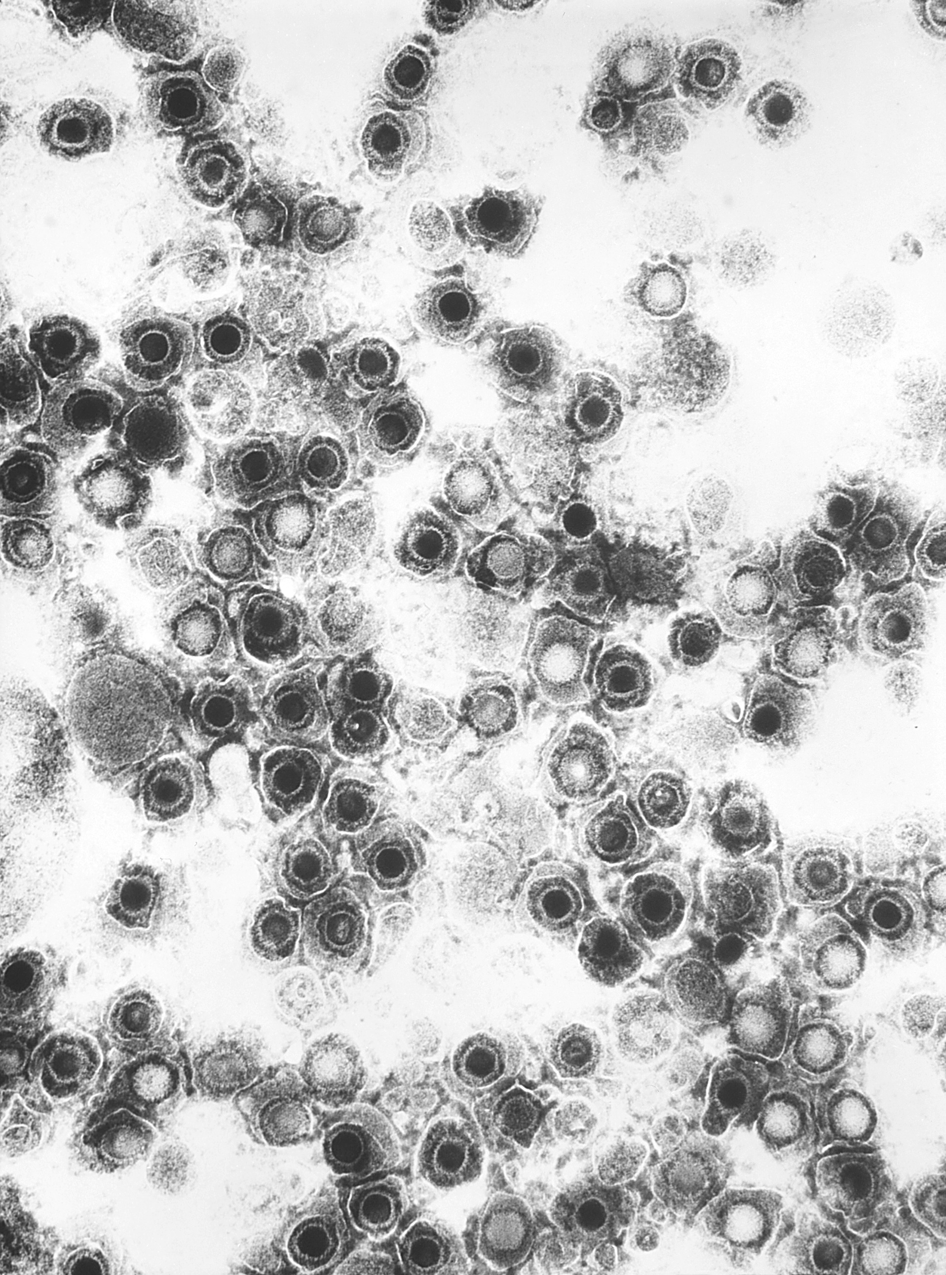
This electron micrograph shows Coxsackie B4 virus particles.
Public domain/ Centers for Disease Control and Prevention/ 1981
http://phil.cdc.gov/phil/imageidsearch.asp (Image ID # 5630)
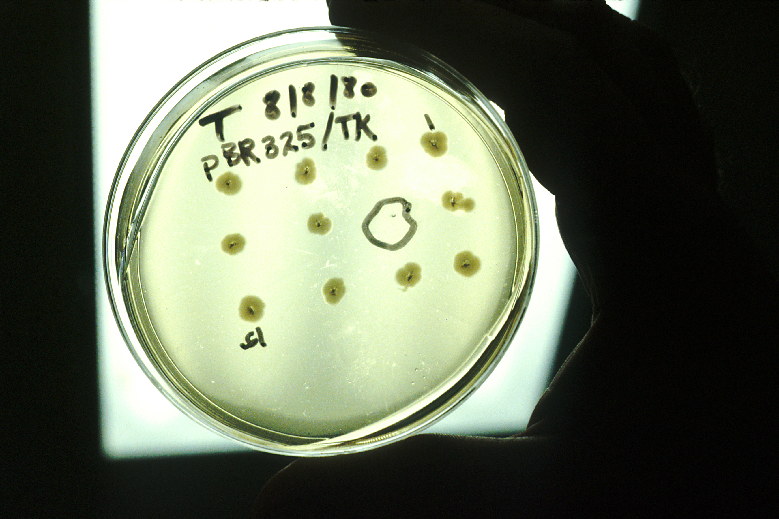
This culture medium shows a gene of herpes simplex virus being cloned in bacteria.
Public domain/ National Cancer Institute/ Linda Bartlett, photographer/ 1980
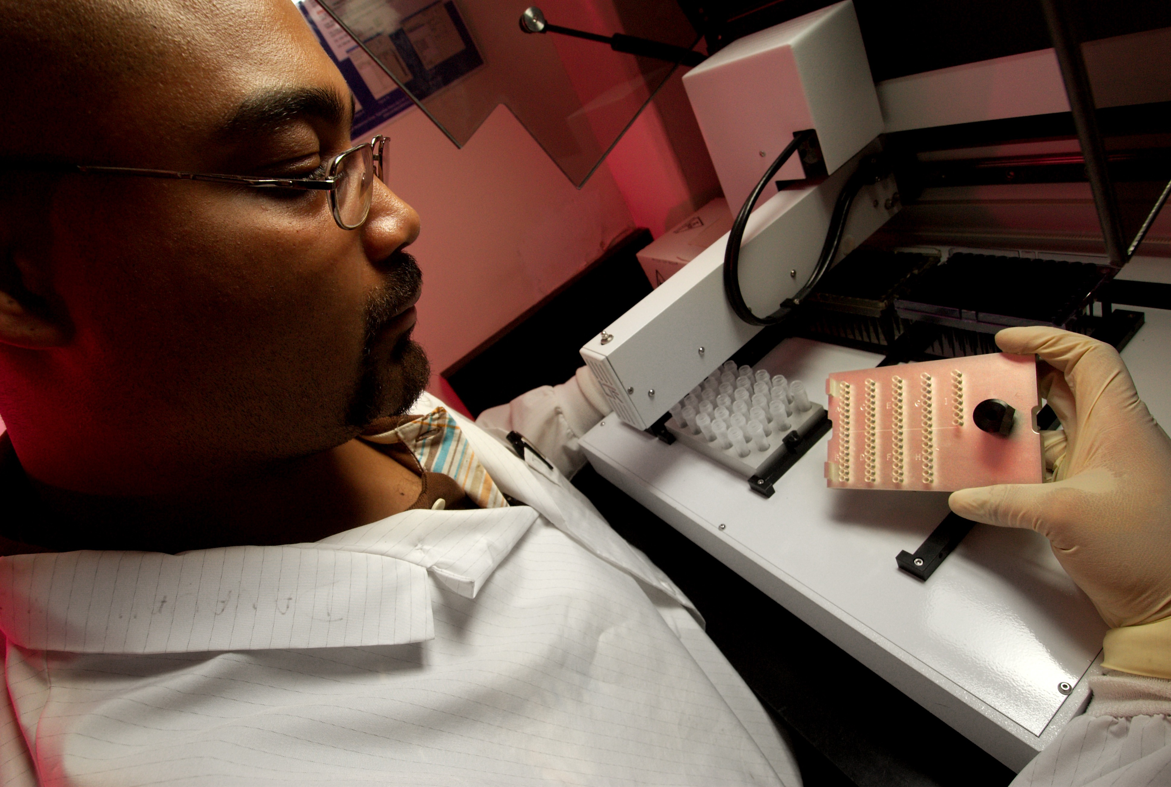
A biologist sets up a liquid-handling robot in order to perform a PCR (polymerase chain reaction) analysis to detect the presence of herpes simplex virus type 2 (HSV-2).
Public domain/ Centers for Disease Control and Prevention/ Hsi Liu, Ph.D., MBA and James Gathany, content providers/ 2007
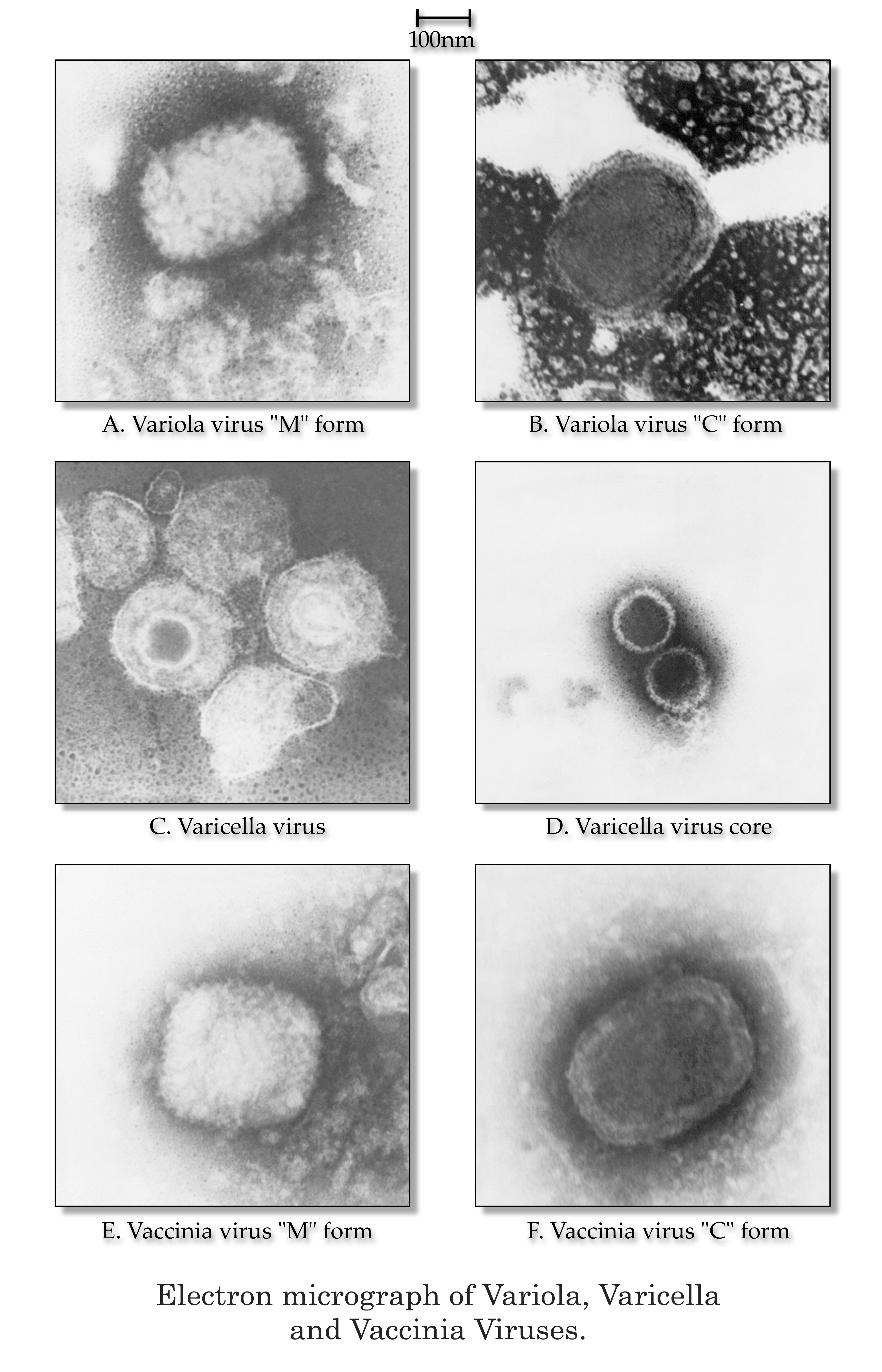
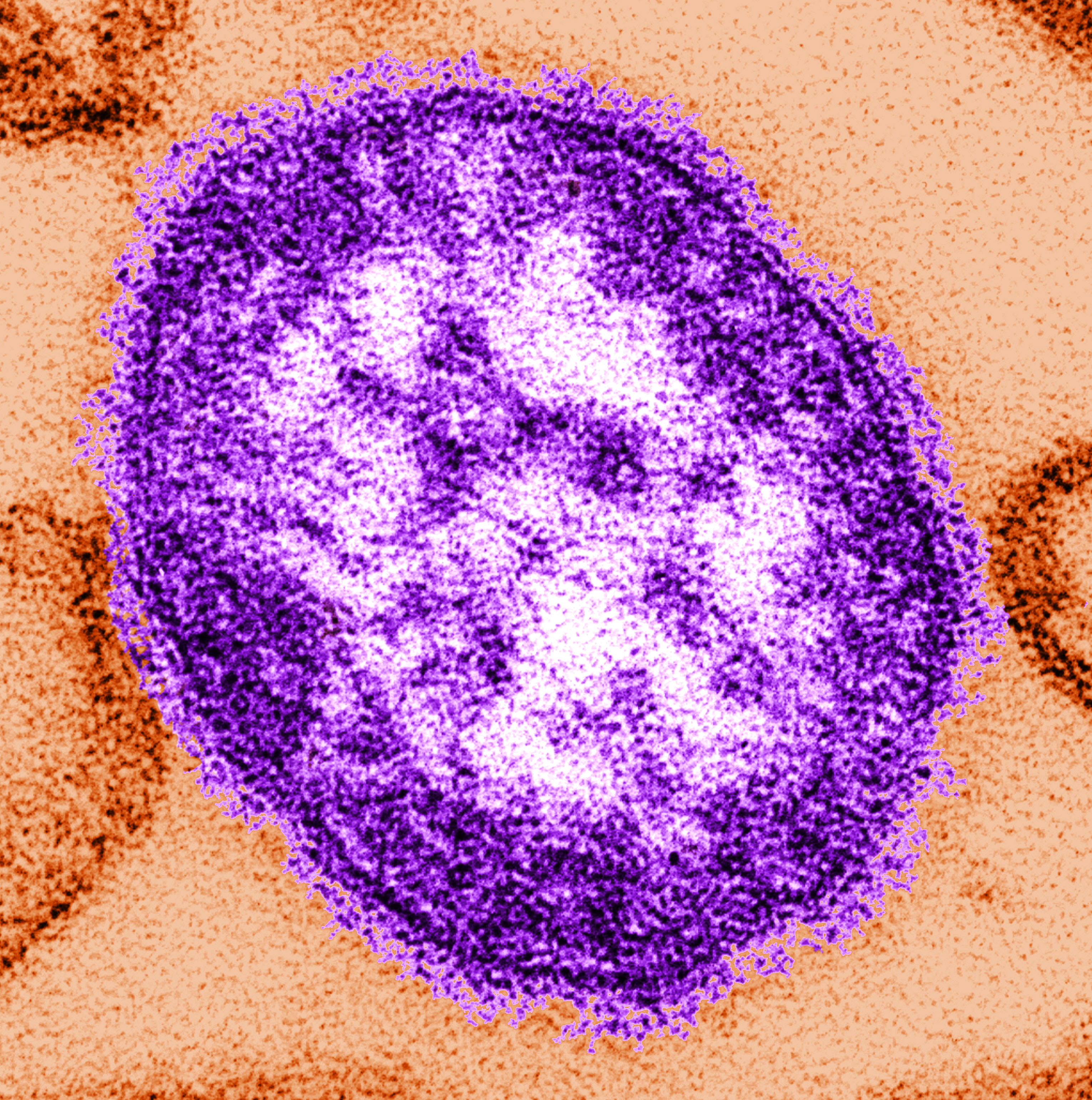
This electron micrograph shows variola, varicella, and vaccinia virions.
Public domain/ Centers for Disease Control and Prevention/ Dr. James Nakano, content provider/ 1975
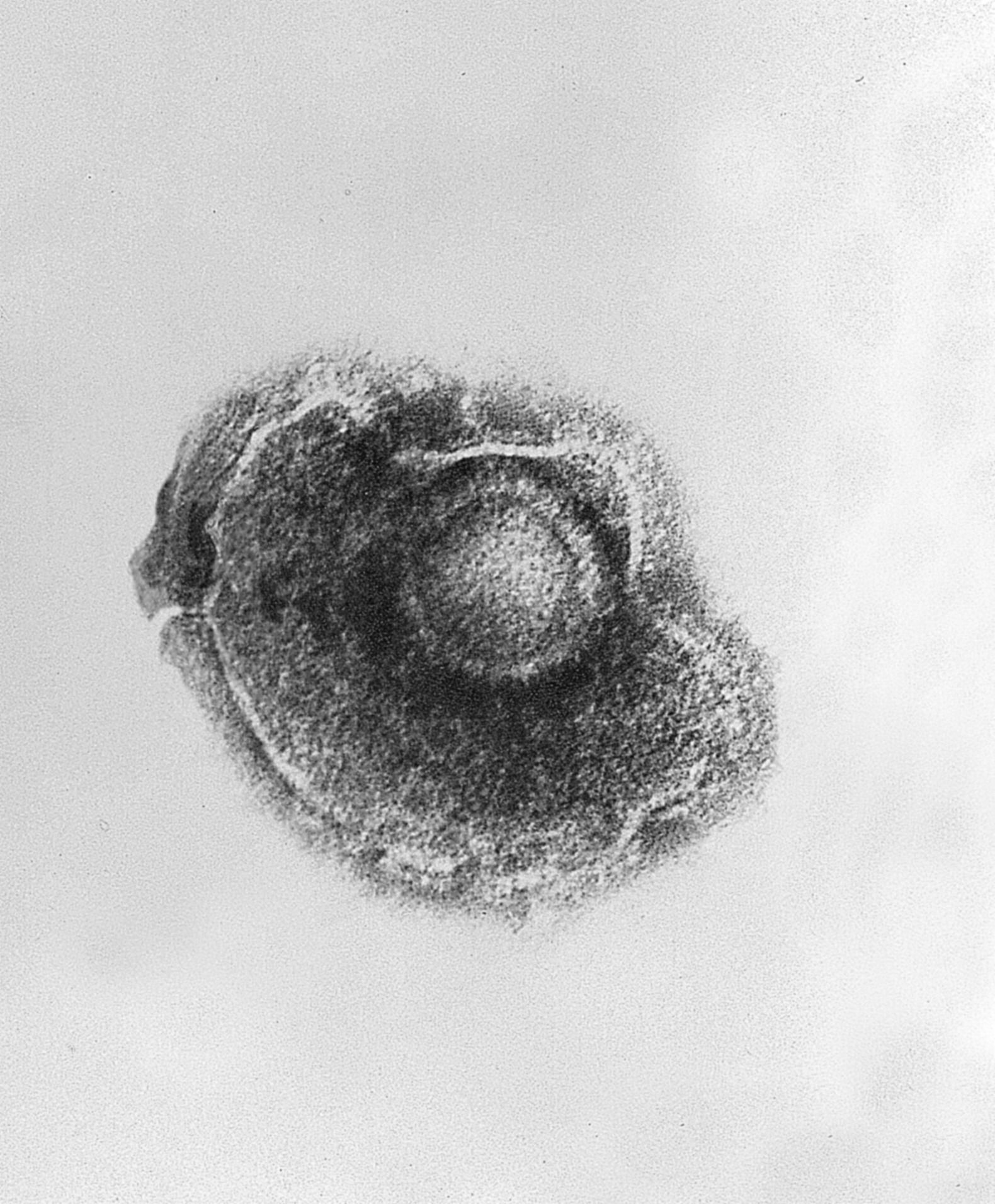
This electron micrograph shows the varicella (chickenpox) virus.
Public domain/ Centers for Disease Control and Prevention/ Dr. Erskine Palmer and B.G. Partin, content providers/ 1982


