Description
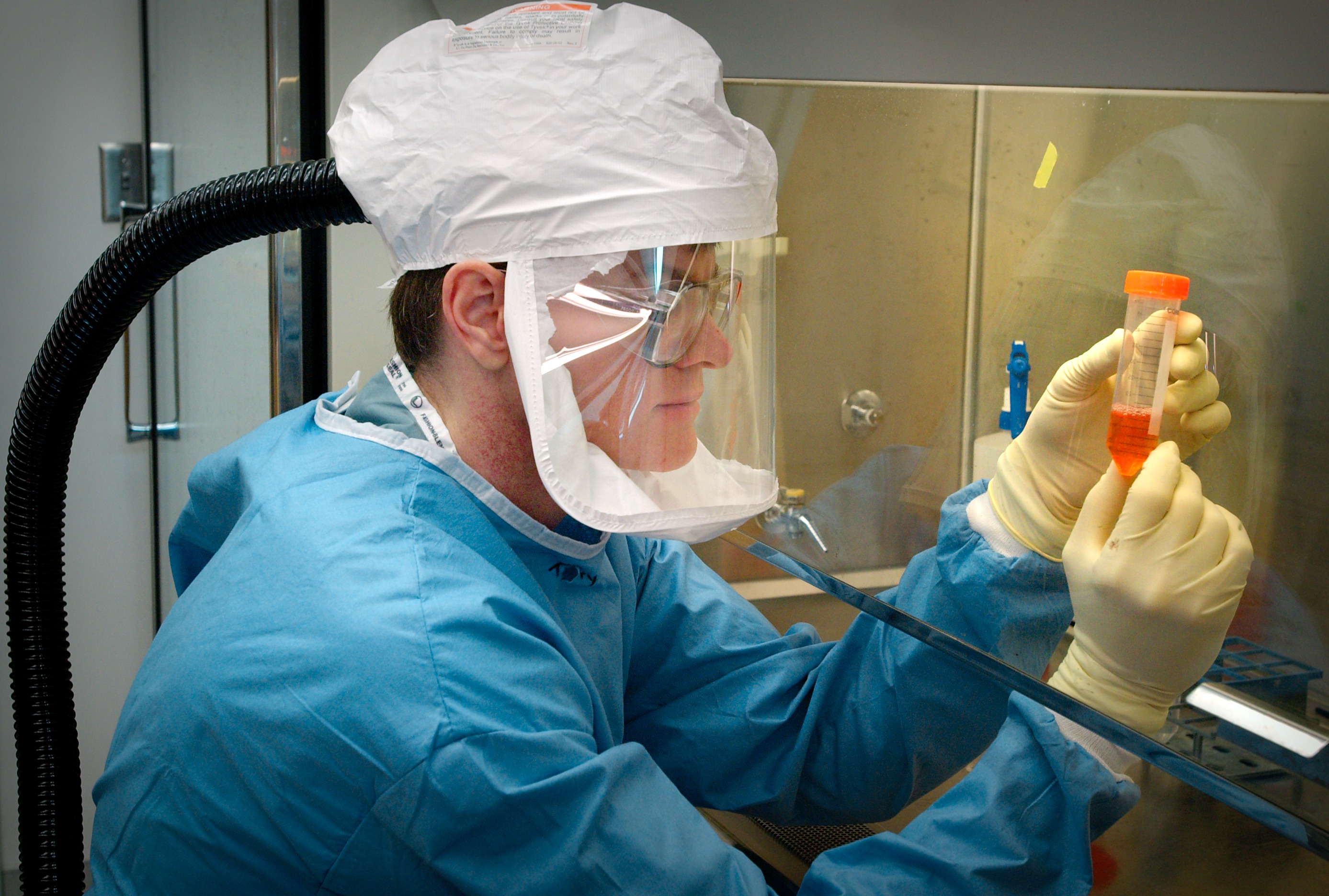
This photograph shows a microbiologist examining a test tube of reconstructed 1918 pandemic influenza viruses.
Credit
Public domain/ Centers for Disease Control and Prevention/ James Gathany, photographer/ 2005
This photograph shows a microbiologist examining a test tube of reconstructed 1918 pandemic influenza viruses.
Public domain/ Centers for Disease Control and Prevention/ James Gathany, photographer/ 2005
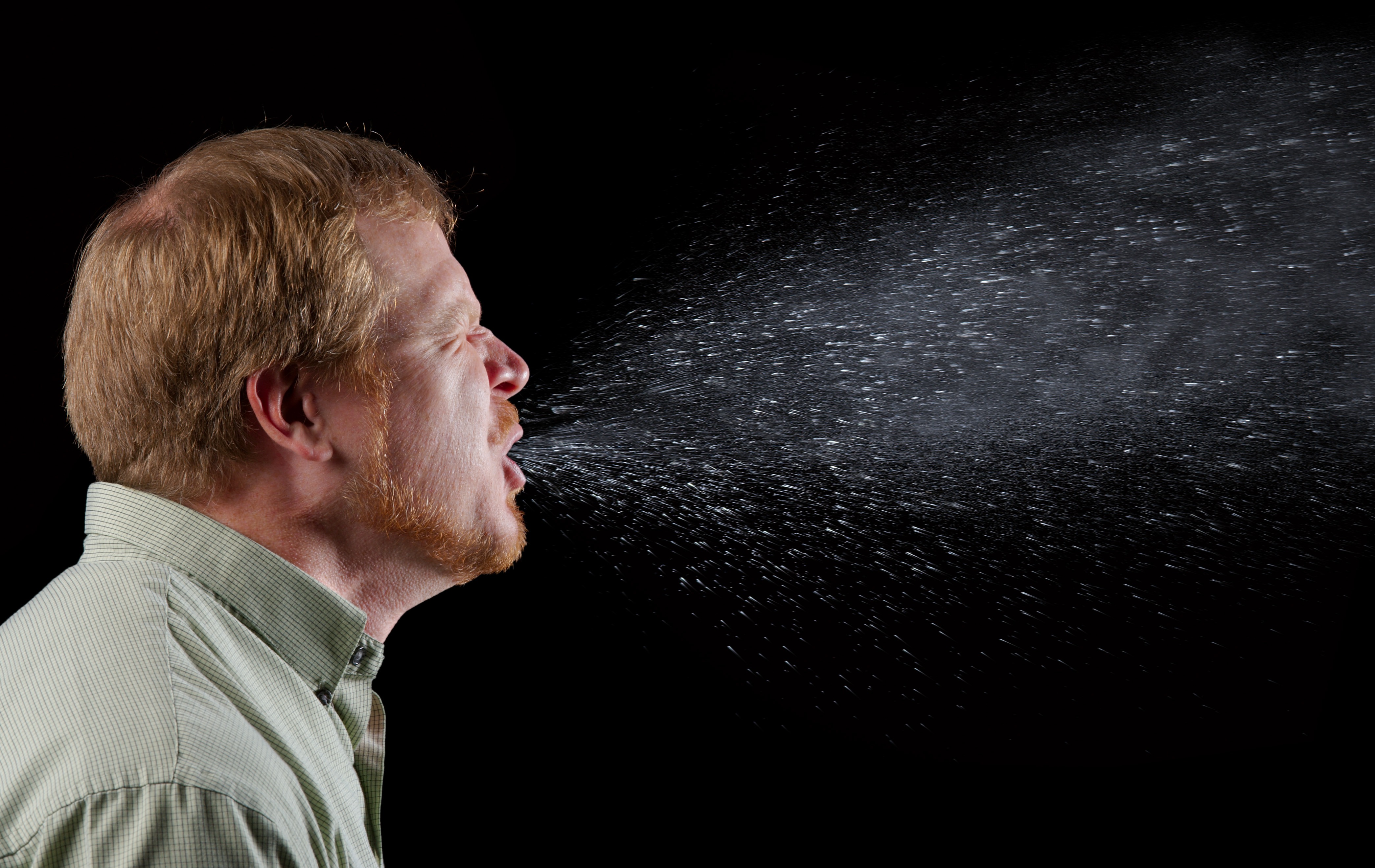
This photograph captures a sneeze in progress. It shows the plume of salivary droplets this man is expelling.
Public domain/ Centers for Disease Control and Prevention/ Brian Judd, content provider/ James Gathany, photographer/ 2009
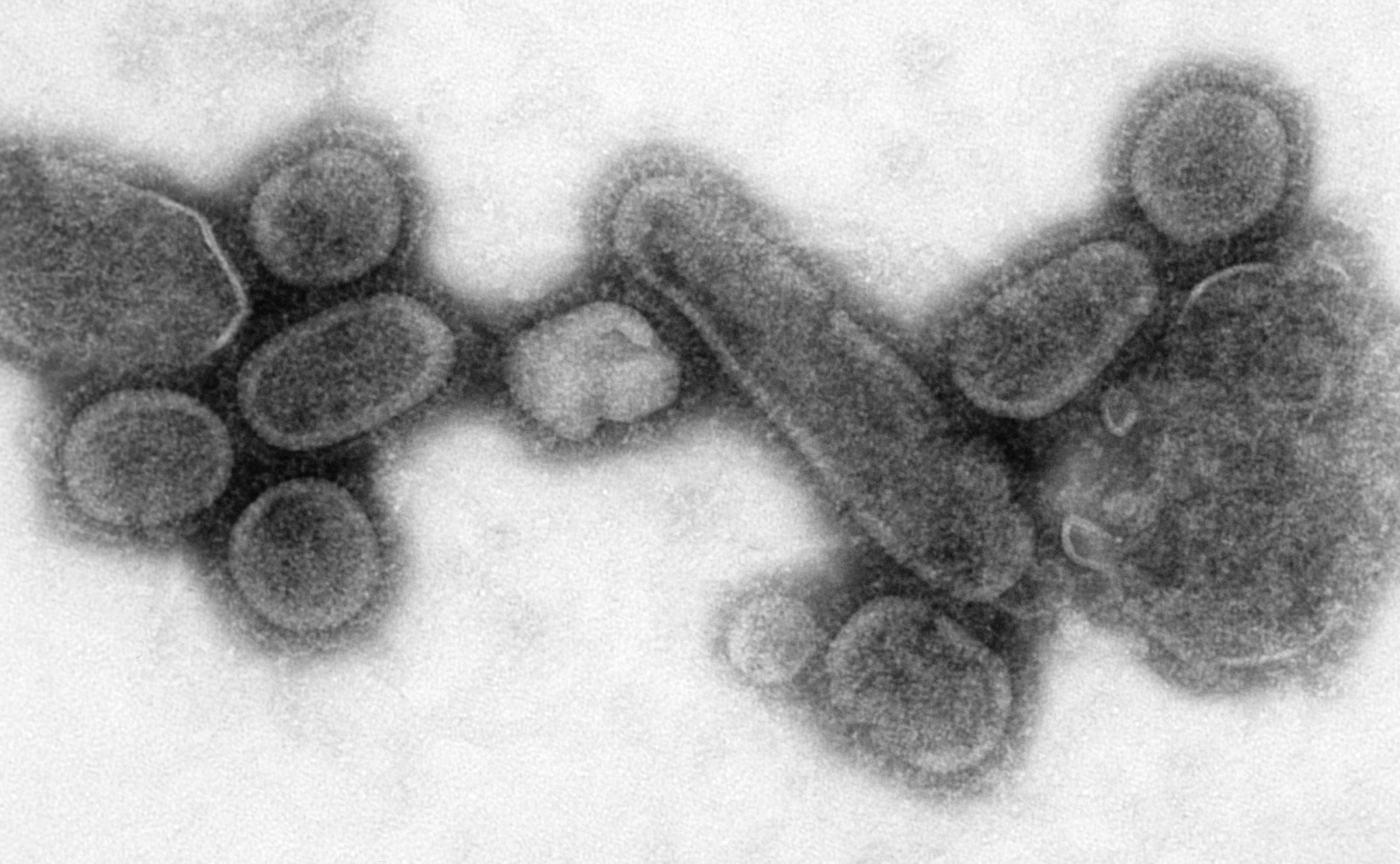
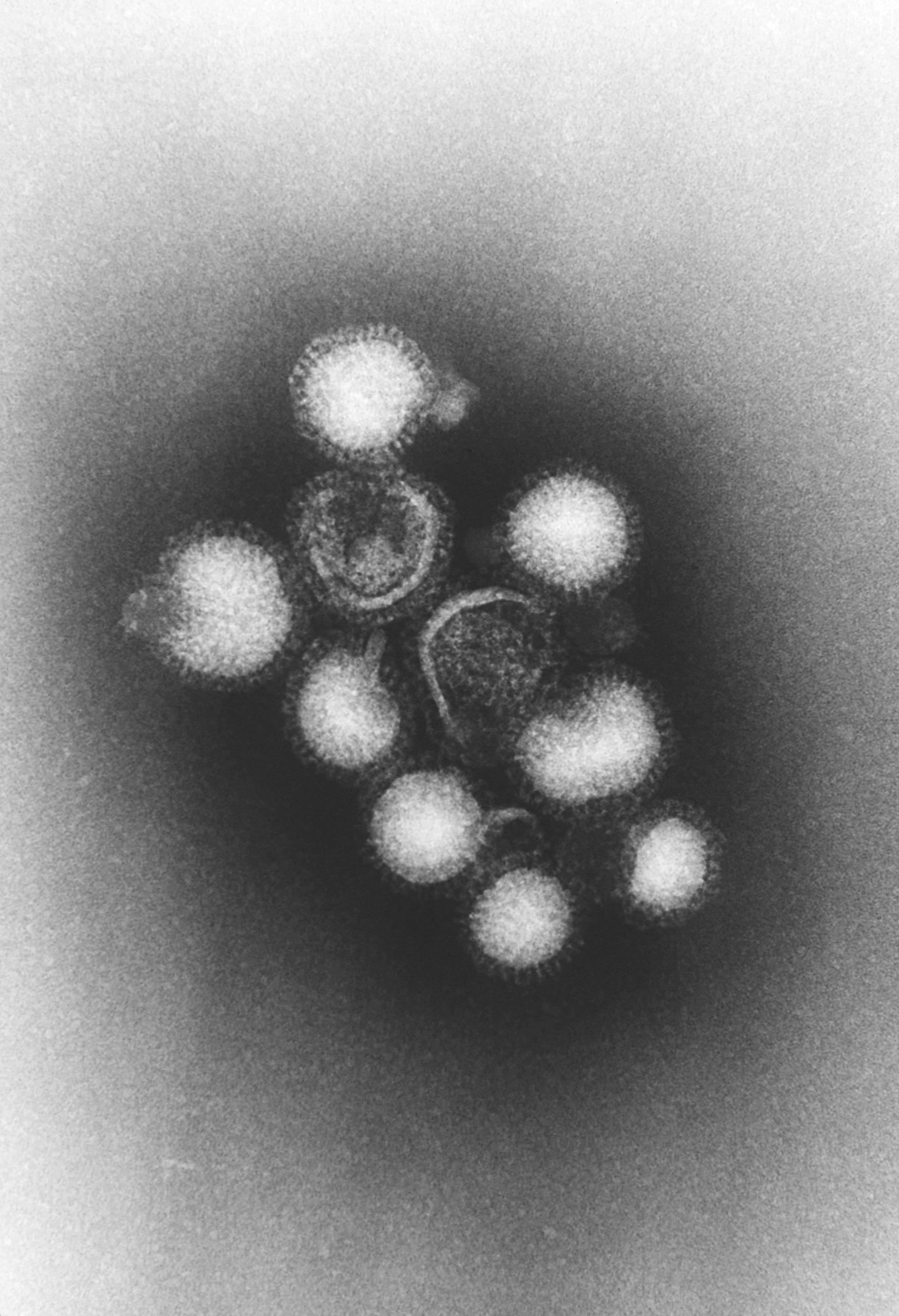
This electron micrograph shows 1918 influenza virions.
Public domain/ Centers for Disease Control and Prevention/ Dr. Terrence Tumpey, content provider/ Cynthia Goldsmith, photographer/ 2005
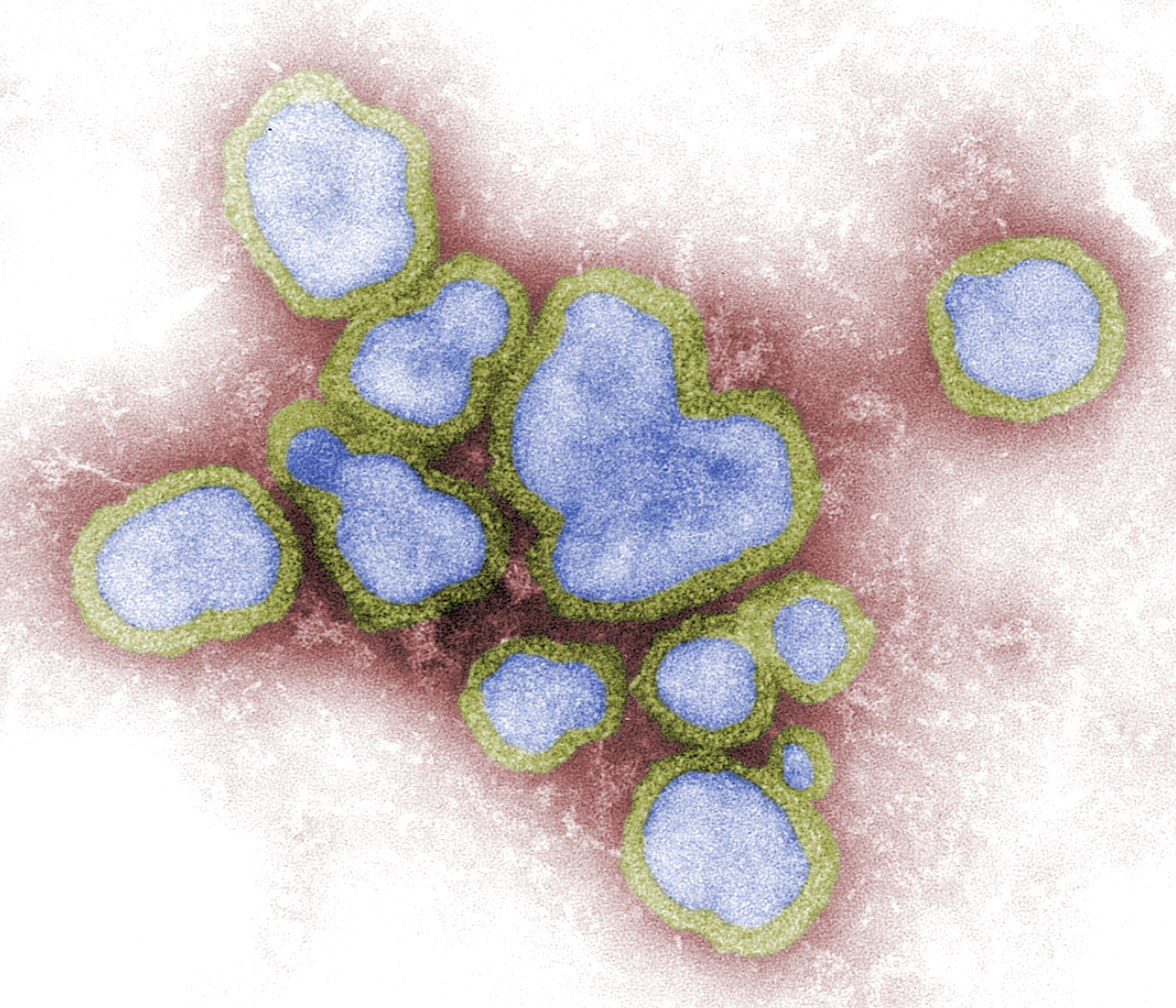
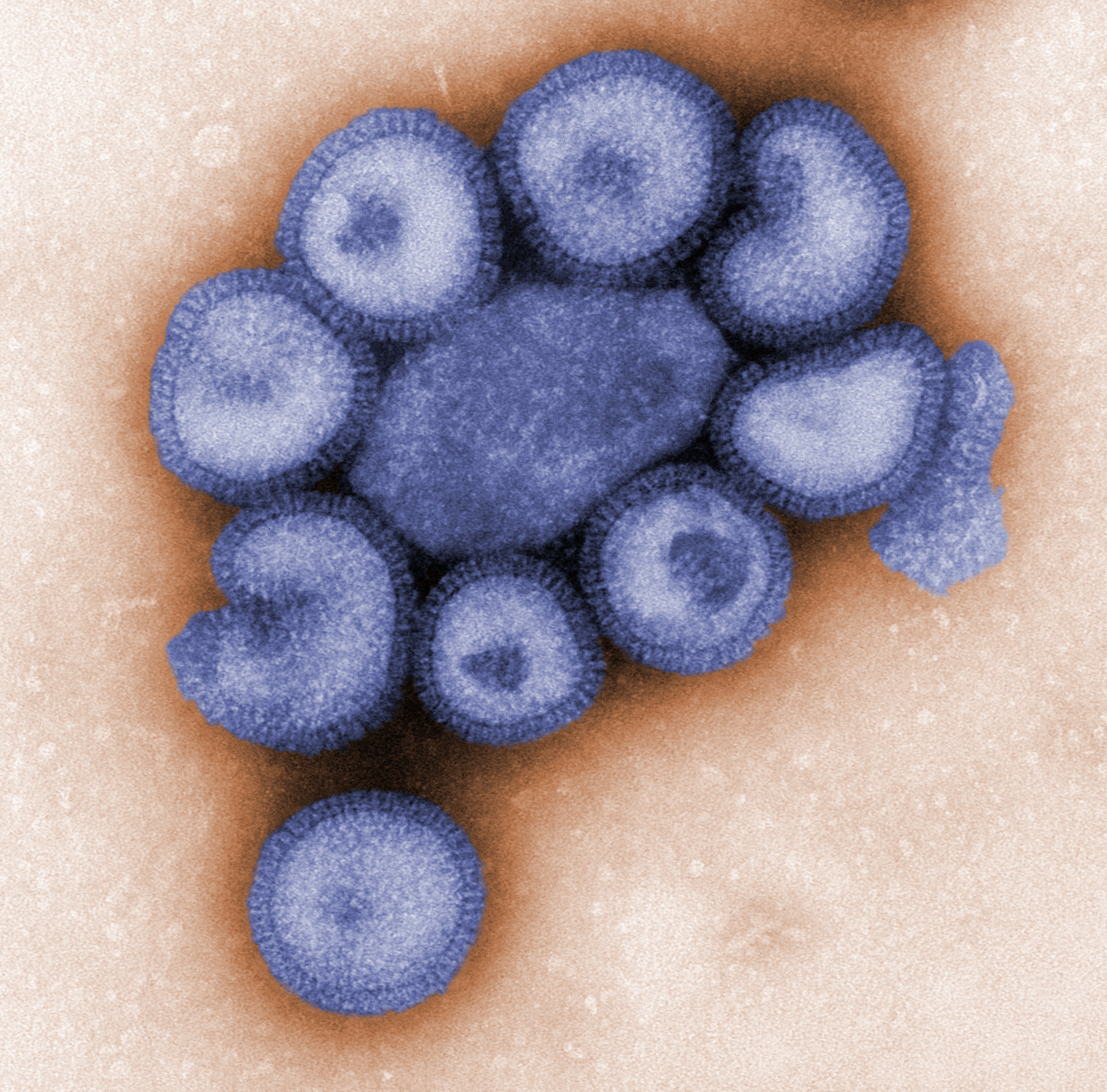
Electron micrograph shows a Hong Kong H3N2 subtype of the influenza A virus.
Public domain/ Centers for Disease Control and Prevention/ Dr. Fred Murphy, content provider/ 1975
This electron micrograph shows a number of influenza virions. Notice the neuraminidase (N) and hemagglutinin (H) spikes sticking out from the protein coat, or capsid.
Public domain/ Centers for Disease Control and Prevention/ Dr. John Hierholzer, content provider/ 1974
This electron micrograph shows an H1N1 influenza virus that causes disease in pigs. It is magnified 37,800 times. Several virus particles are developing while being grown in chicken eggs.
Public domain/ Centers for Disease Control and Prevention/ Dr. E. Palmer and R.E. Bates, content providers/ 1976
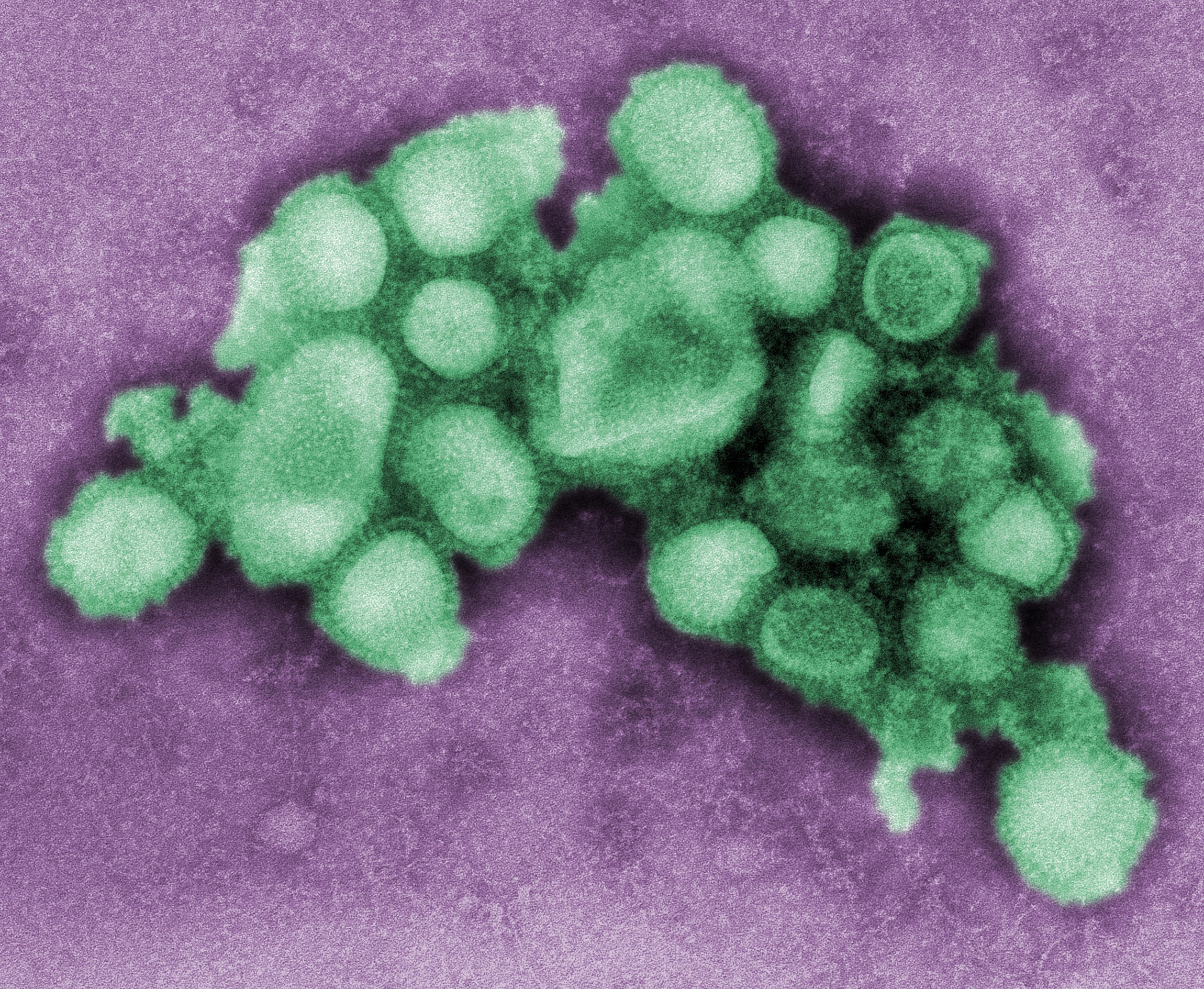
This electron micrograph shows virus particles, or virions, from Type A influenza virus.
Public domain/ Centers for Disease Control and Prevention/ F.A. Murphy, content provider/ 1976
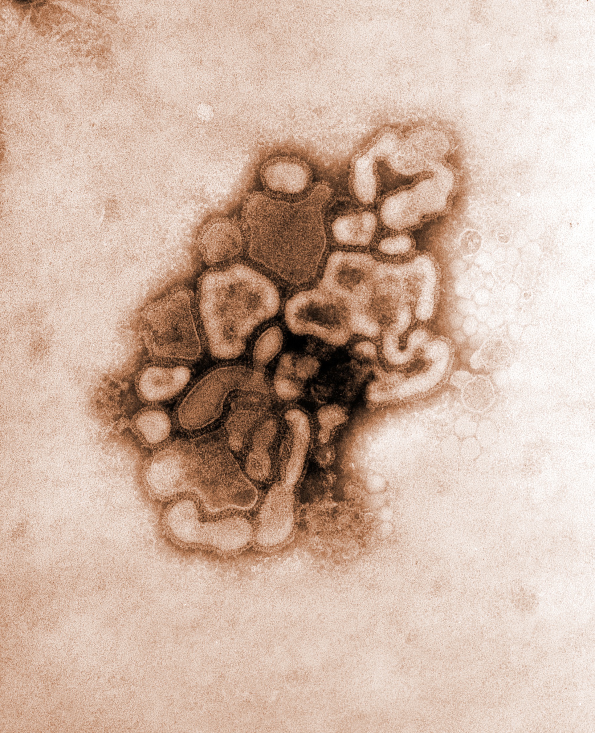
This electron micrograph shows several virus particles, or virions, of a re-created 1918 influenza virus growing in kidney cells of a dog.
Public domain/ Centers for Disease Control and Prevention/ Dr. Terrence Tumpey, content provider/ Cynthia Goldsmith, photographer/ 2005
Electron micrograph shows a number of influenza virus particles, or virions.
Public domain/ Centers for Disease Control and Prevention/ Dr. F. A. Murphy, content provider/ 1973
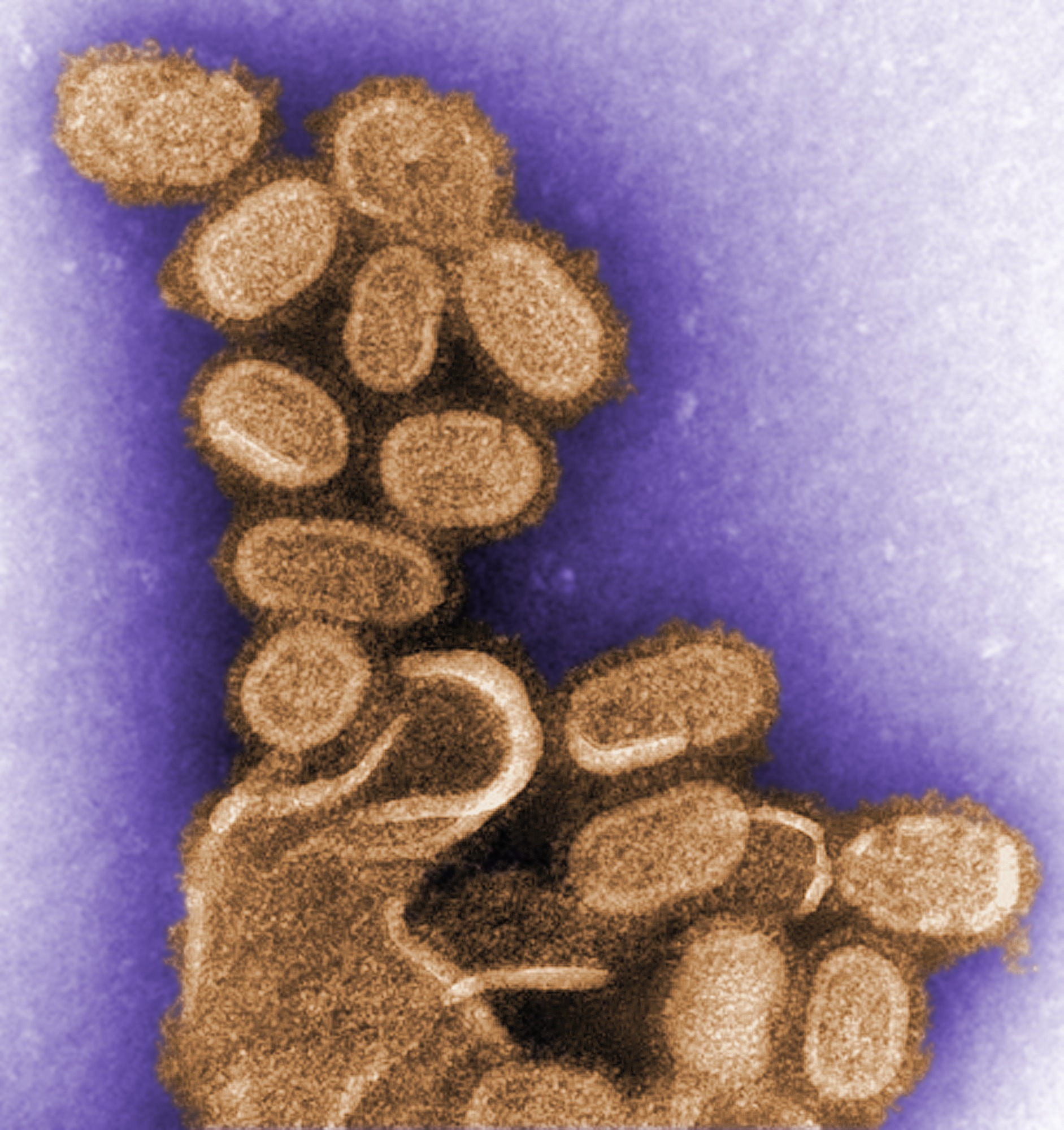
This electron micrograph shows several particles, or virions, of an influenza virus that causes the disease in pigs.
Public domain/ Centers for Disease Control and Prevention/ C. S. Goldsmith and A. Balish, content providers/ 2009