CONTACT

WOV

  • COVID-19
    • C’RONA COMIX
    • TRIBAL C’RONA COMIX
    • C’RONA COMIX II
    • C’RONA PANDEMIC COMICS
  • COMICS & Apps
    • C’RONA PANDEMIC COMICS
    • CARNIVAL OF CONTAGION
    • Confined!
    • Phantom Planet
    • The Never-Ending Battle
    • The Frozen Horror
    • Curse of the Tree-man
    • Occupied by microbes!
    • Microbe Stickers
  • Books & Audio
    • C’RONA PANDEMIC COMICS
    • PLANET OF VIRUSES
    • World of Viruses
    • Watch Your Mouth!
    • Microbe Stickers
    • CARNIVAL OF CONTAGION
    • Audio Stories
  • Microbe Images
    • Inside Viruses
    • Microbe Scientists
    • Microbe SEMS
    • Historical Images
    • Microbe Illustrations
  • Activities
  • Omaha Sci Media
  • Worlds of Connections
  • Research & Evaluation

Image wall

Leukemia cells containing Epstein-Barr virus

January 9, 2013 jbrehm2
Description

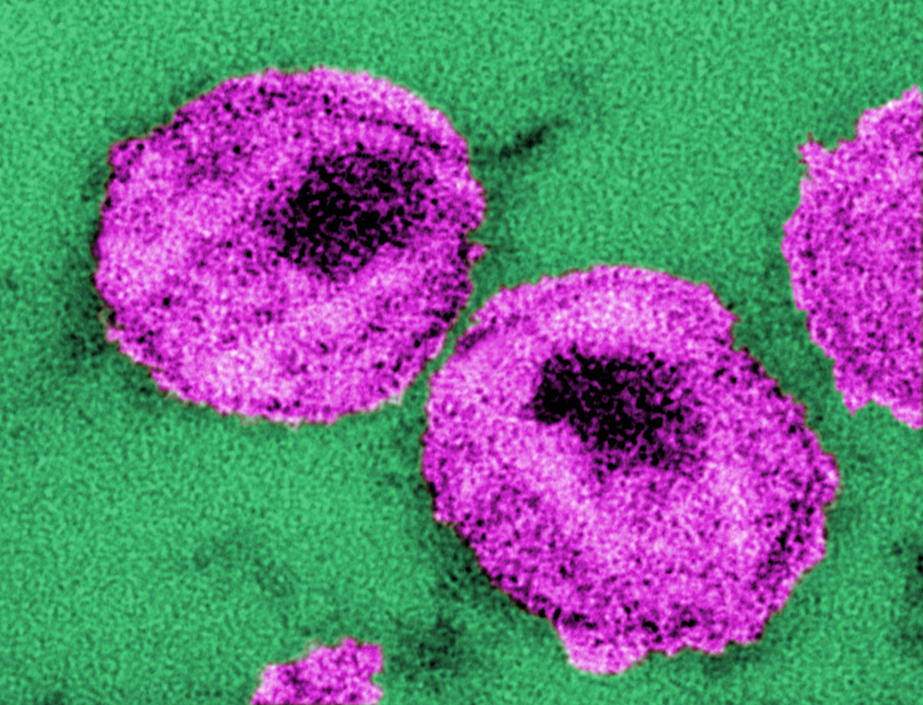
This electron micrograph shows leukemia cells (cancerous white blood cells) that contain Epstein-Barr virus.

Credit

Public domain/ Centers for Disease Control and Prevention/ Dr. Paul M. Feorino, content provider/ 1972

Source

http://phil.cdc.gov/phil/imageidsearch.asp (Image ID # 2984)

Image wall

Electron micrograph of HIV detailed structures

January 9, 2013 jbrehm2
Description

This electron micrographic image shows the structures of HIV particles.

Credit

Public domain/ Centers for Disease Control and Prevention/ Dr. A. Harrison and Dr. P. Feorino, content providers/ Unknown date

Source

http://phil.cdc.gov/phil/imageidsearch.asp (Image ID # 10860)

Image wall, Microbe SEMS

Electron micrograph of HTLV-1 and HIV

January 9, 2013 jbrehm2
Description

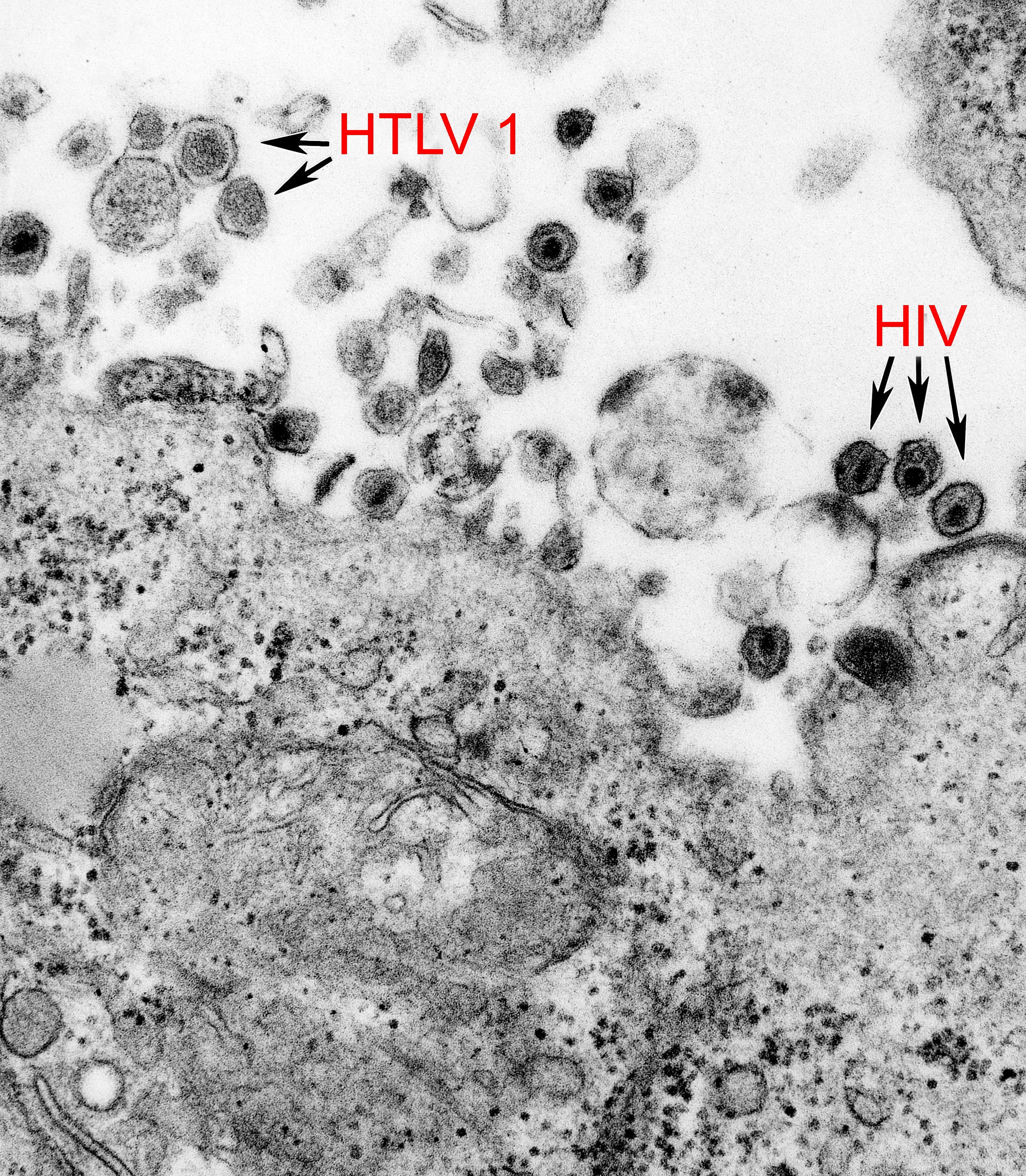
This electron micrograph shows both the human T-cell leukemia type-1 virus (HTLV-1) and the human immunodeficiency virus (HIV).

Credit

Public domain/ Centers for Disease Control and Prevention/ Cynthia Goldsmith, content provider/ Unknown date

Source

http://phil.cdc.gov/phil/imageidsearch.asp (Image ID # 8241)

Image wall, Microbe SEMS

Electron micrograph of HIV mature form

January 9, 2013 jbrehm2
Description

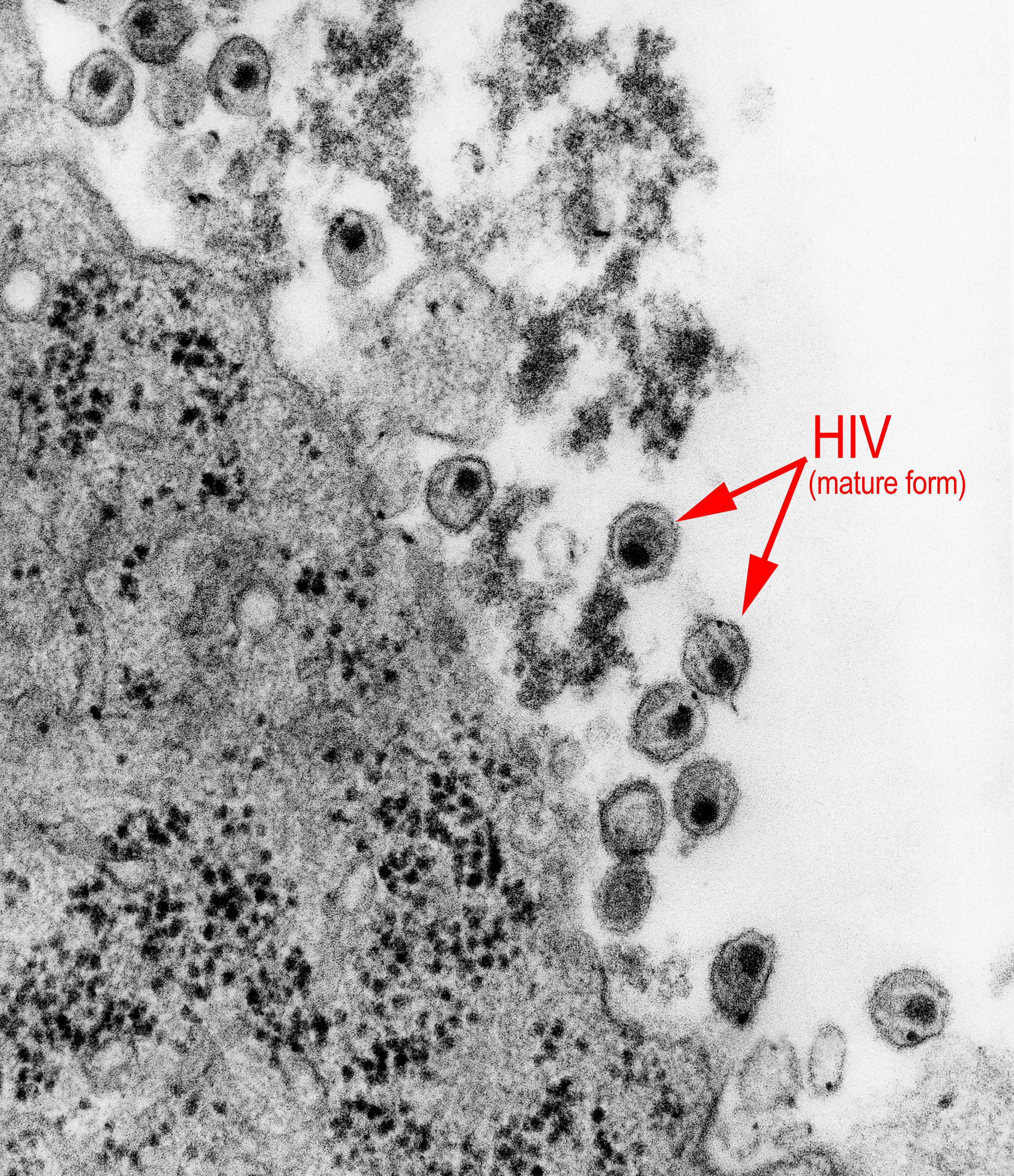
This electron micrograph shows mature forms of the human immunodeficiency virus (HIV).

Credit

Public domain/ Centers for Disease Control and Prevention/1983

Source

http://phil.cdc.gov/phil/imageidsearch.asp (Image ID # 8254)

Image wall, Microbe Illustrations

HIV entering a cell

January 9, 2013 jbrehm2
Description

This illustration shows HIV entering a cell and releasing its genetic material (RNA) and proteins inside the cell.

Credit

Public domain/ National Institutes of Health/ Unknown date

Source

http://history.nih.gov/NIHInOwnWords/docs/page_40a.html

Image wall, Microbe SEMS

HIV “budding” out of a human immune cell

January 9, 2013 jbrehm2
Description

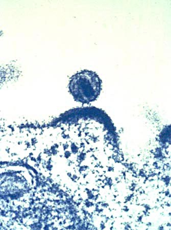
This electron photomicrograph shows HIV budding out of a human immune cell, which the virus infects and uses to make copies.

Credit

Public domain/ National Institute of Allergy and Infectious Diseases, National Institutes of Health/ courtesy of Dr. Tom Folks/ 2008

Source

http://history.nih.gov/NIHInOwnWords/docs/page_40b.html

Image wall, Microbe Illustrations

Diagram of HIV reproduction

January 9, 2013 jbrehm2
Description

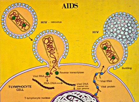
This illustration shows how HIV attaches to an immune cell and makes copies of itself.

Credit

Public domain/ National Institutes of Health/ Unknown date

Source

http://history.nih.gov/NIHInOwnWords/docs/page_40c.html

Image wall, Microbe SEMS

HIV-infected T cells

January 9, 2013 jbrehm2
Description

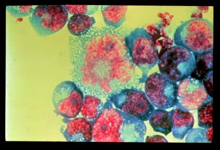
This electron photomicrograph shows T cells infected with HIV.

Credit

Public domain / National Institute of Allergy and Infectious Diseases, National Institutes of Health / Department of Health and Human Services, courtesy of Dr. Tom Folks / 2008

Source

http://media.nih.gov/imagebank/display_search.aspx?id=232

Image wall, Microbe Illustrations

HIV-1 virion

January 9, 2013 jbrehm2
Description

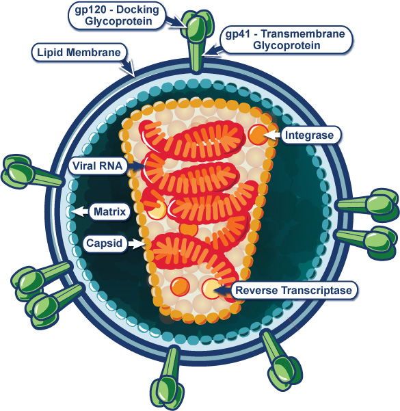
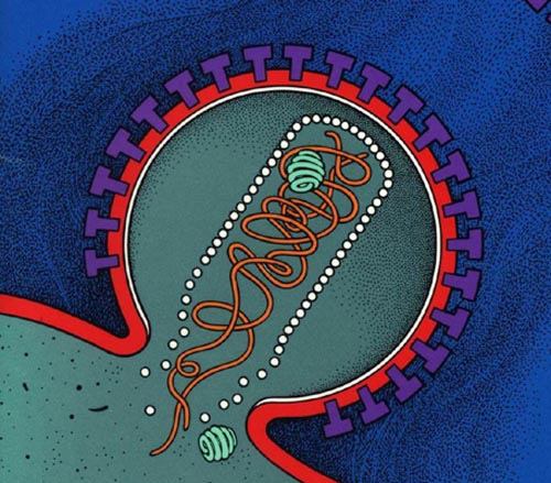
This illustration of HIV structure shows the envelope, capsid, two RNA strands, and outer and inner proteins.

Credit

Public domain/ National Institute of Allergy and Infectious Diseases, National Institutes of Health/ 2009

Source

http://www.niaid.nih.gov/topics/HIVAIDS/Understanding/Biology/Pages/hivVirionLargeImage.aspx

Image wall

Electron micrograph of HIV

January 9, 2013 jbrehm2
Description

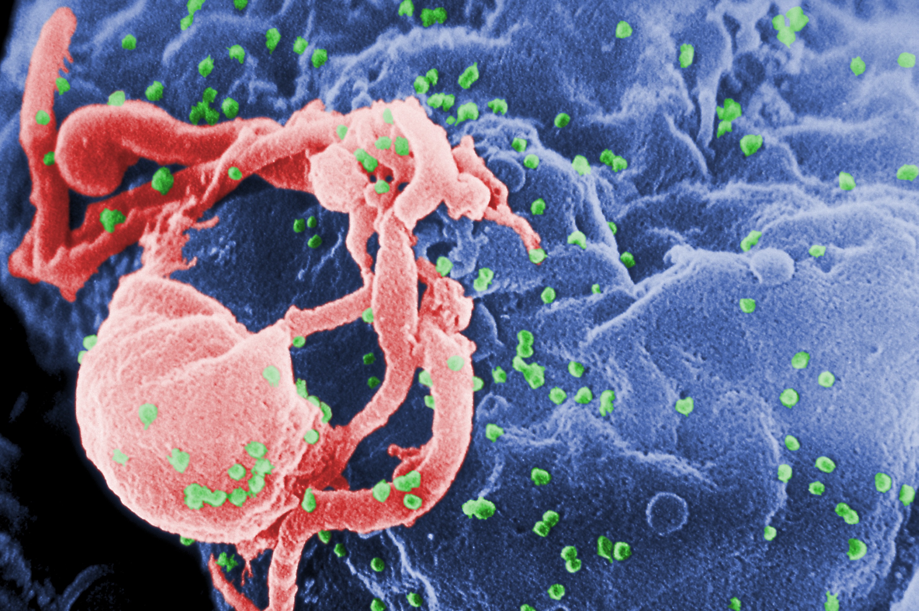
This electron photomicrograph shows HIV (in green) budding from a white blood cell.

Credit

Public domain/ Centers for Disease Control and Prevention/ C. Goldsmith, P. Feorino, E. L. Palmer, and W. R. McManus, content providers/ C. Goldsmith, photographer/ Unknown date

Source

http://phil.cdc.gov/phil/imageidsearch.asp (Image ID # 10000)

Posts navigation

← Previous 1 2 3 4 … 18 Next →

Recent Posts

  • Tribal C’rona Comix Page 5
  • Tribal C’rona Comix Page 4
  • Tribal C’rona Comix Page 3
  • Tribal C’rona Comix Page 2
  • Tribal C’rona Comix Page 1

Recent Comments

    Archives

    • December 2020
    • November 2020
    • October 2020
    • September 2020
    • August 2020
    • July 2020
    • June 2020
    • February 2015
    • December 2014
    • January 2013

    Categories

    • Apps and Activities
    • Bacterial Viruses
    • Covid-19
    • Crona Comix II
    • Curricula
    • Dengue Virus
    • Ebola Virus
    • Epidemic Simulations
    • Foot and Mouth Disease Virus
    • General
    • Hantavirus
    • Hepatitis B Virus
    • Historical Images
    • HIV
    • HPV
    • Image wall
    • Influenza Virus
    • Inside Viruses
    • Microbe Illustrations
    • Microbe Scientists
    • Microbe SEMS
    • Polio Virus
    • Rabies Virus
    • SARS Virus
    • Tribal Crona Comix
    • Uncategorized
    • West Nile Virus

    Meta

    • Log in
    • Entries feed
    • Comments feed
    • WordPress.org