CONTACT

WOV

  • COVID-19
    • C’RONA COMIX
    • TRIBAL C’RONA COMIX
    • C’RONA COMIX II
    • C’RONA PANDEMIC COMICS
  • COMICS & Apps
    • C’RONA PANDEMIC COMICS
    • CARNIVAL OF CONTAGION
    • Confined!
    • Phantom Planet
    • The Never-Ending Battle
    • The Frozen Horror
    • Curse of the Tree-man
    • Occupied by microbes!
    • Microbe Stickers
  • Books & Audio
    • C’RONA PANDEMIC COMICS
    • PLANET OF VIRUSES
    • World of Viruses
    • Watch Your Mouth!
    • Microbe Stickers
    • CARNIVAL OF CONTAGION
    • Audio Stories
  • Microbe Images
    • Inside Viruses
    • Microbe Scientists
    • Microbe SEMS
    • Historical Images
    • Microbe Illustrations
  • Activities
  • Omaha Sci Media
  • Worlds of Connections
  • Research & Evaluation

Image wall, Microbe SEMS

Herpes viruses

January 9, 2013 jbrehm2
Description

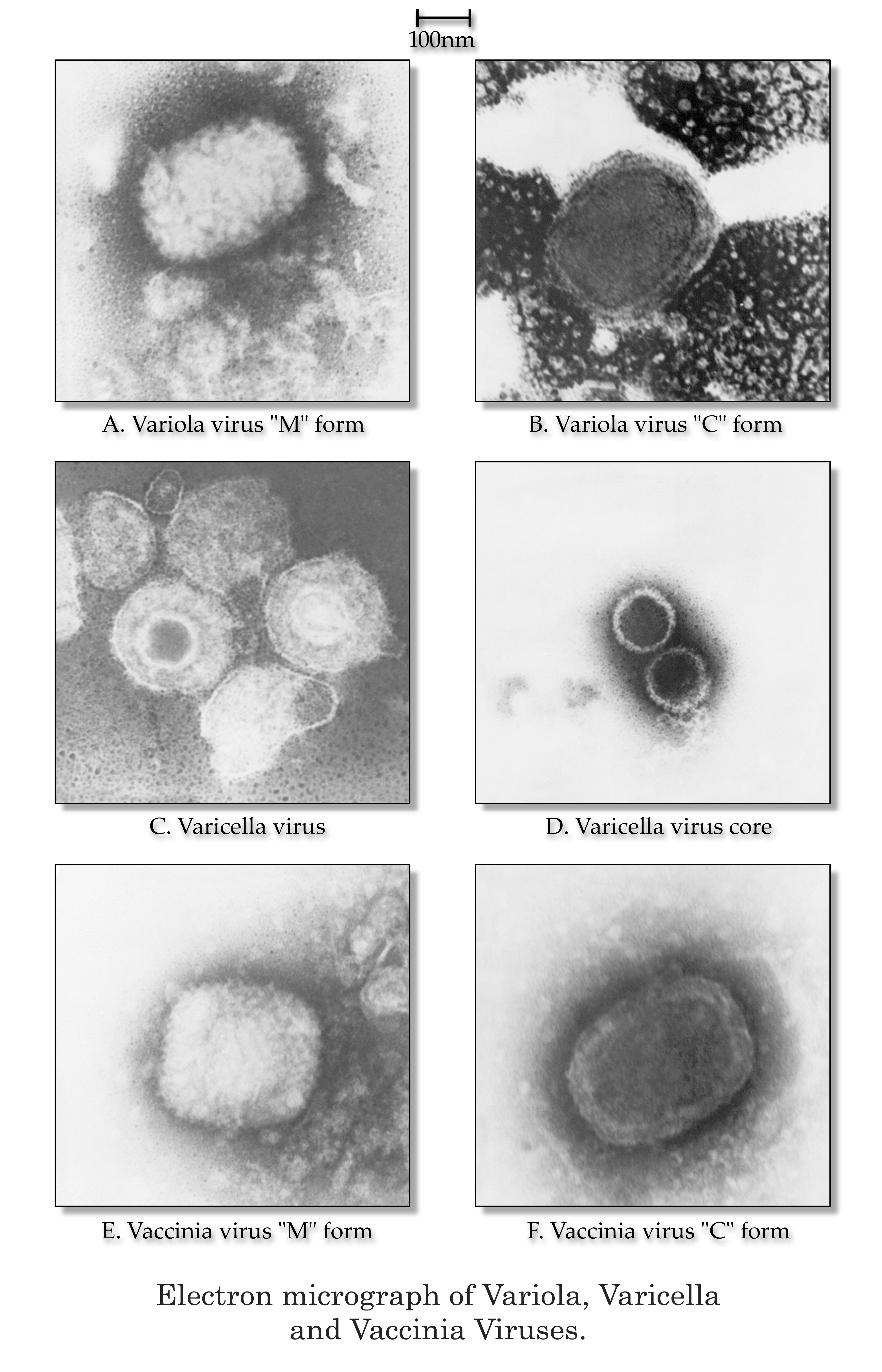
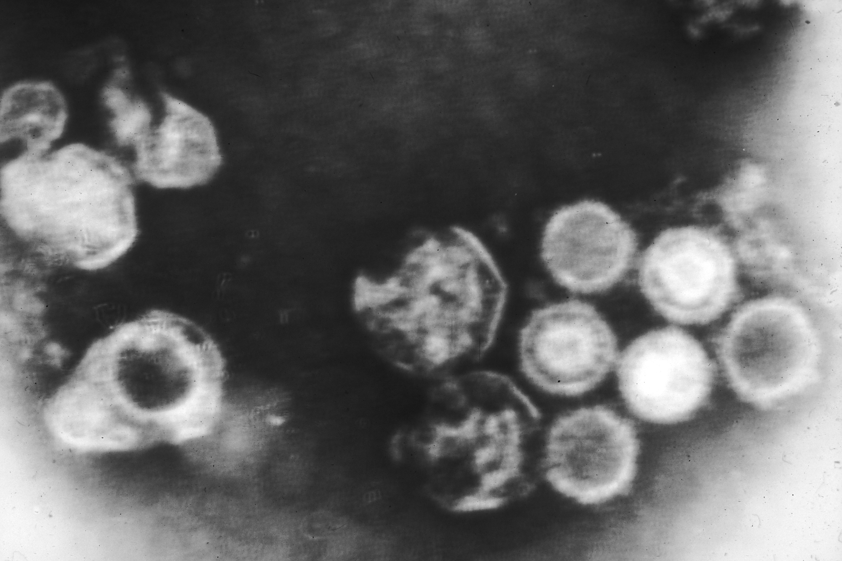
This electron micrograph shows variola, varicella, and vaccinia virions.

Credit

Public domain/ Centers for Disease Control and Prevention/ Dr. James Nakano, content provider/ 1975

Source

http://phil.cdc.gov/phil/imageidsearch.asp (Image ID # 2426)

Image wall, Microbe SEMS

Varicella (chickenpox) virus

January 9, 2013 jbrehm2
Description

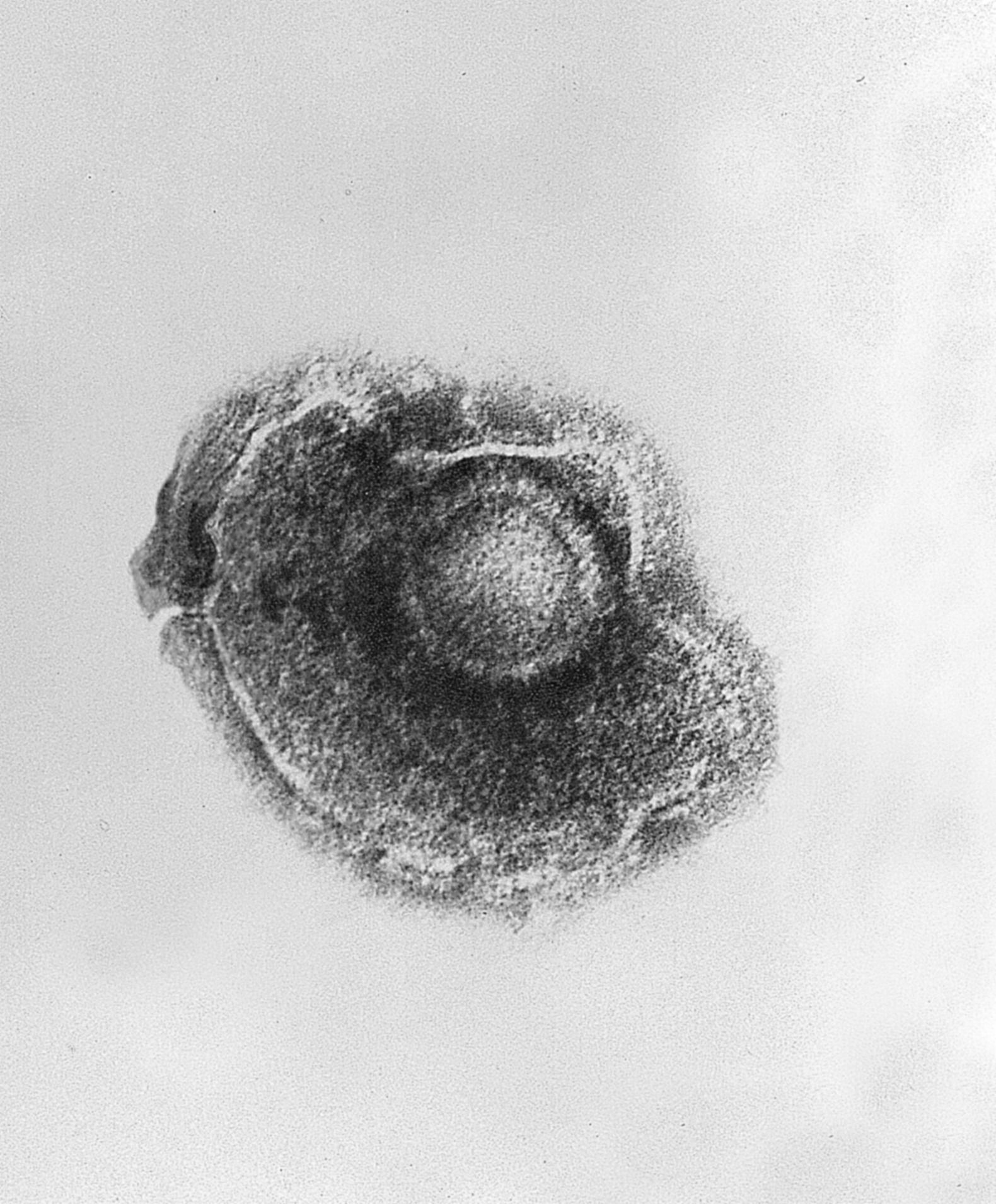
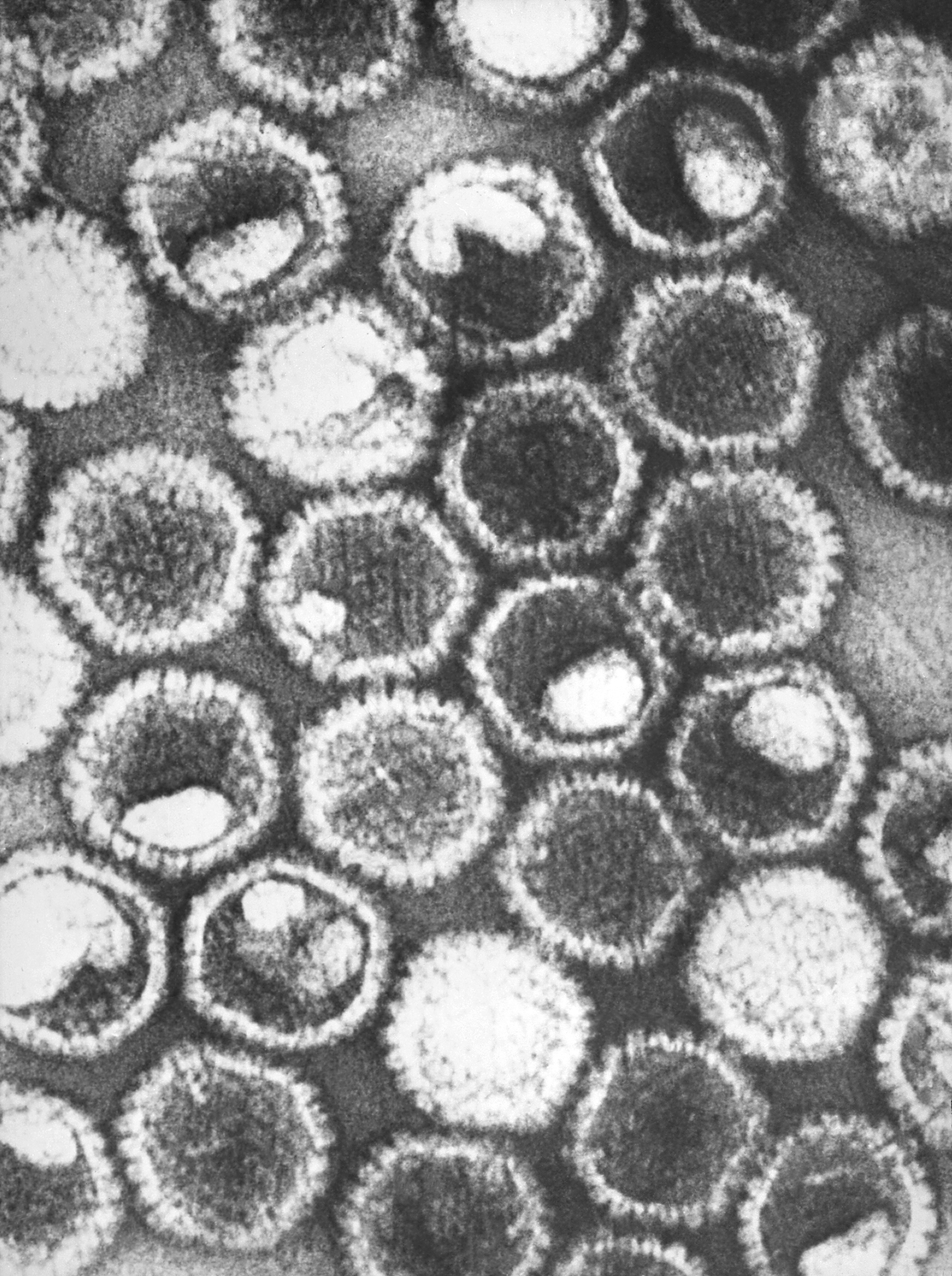
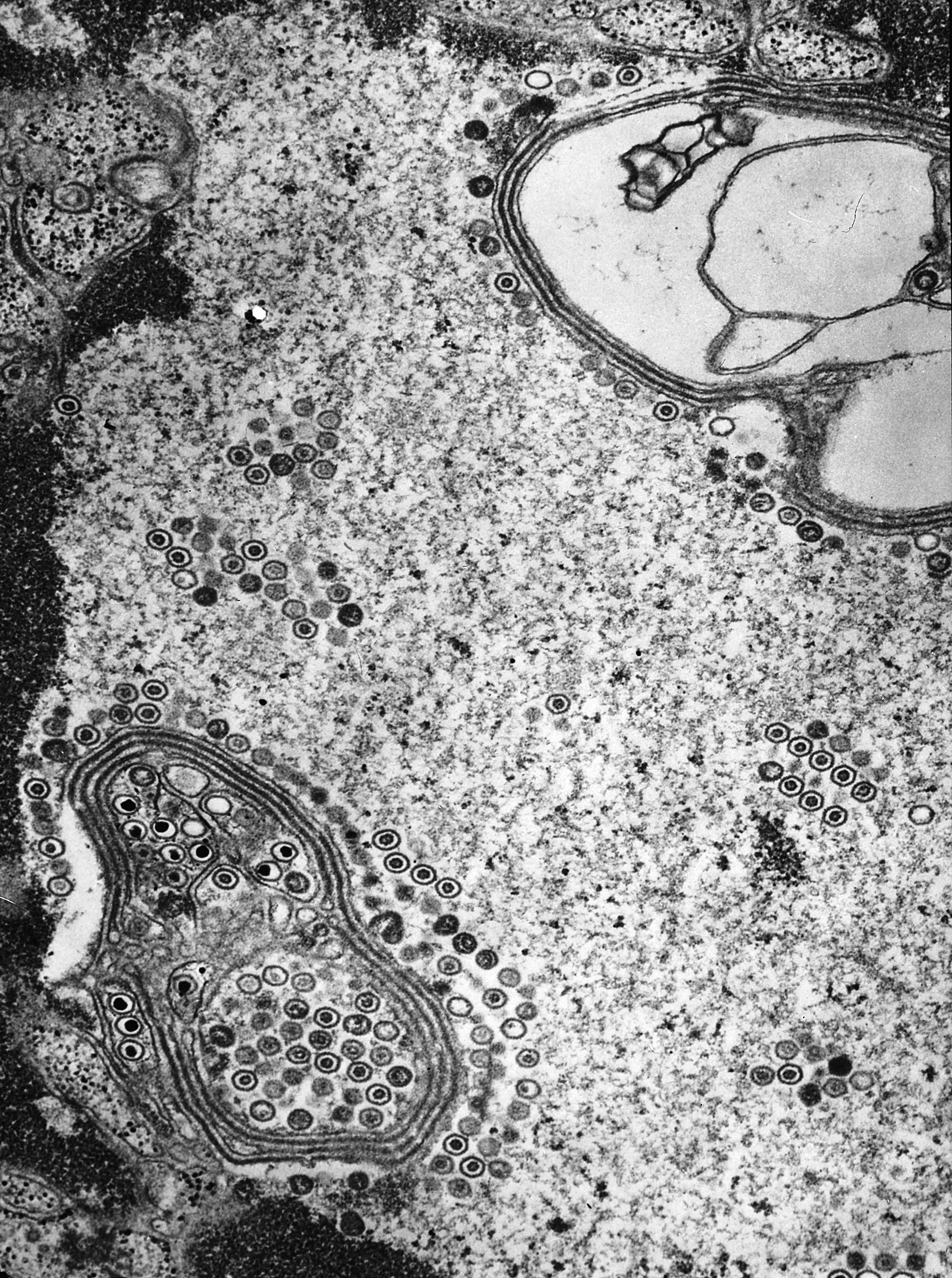
This electron micrograph shows the varicella (chickenpox) virus.

Credit

Public domain/ Centers for Disease Control and Prevention/ Dr. Erskine Palmer and B.G. Partin, content providers/ 1982

Source

http://phil.cdc.gov/phil/imageidsearch.asp (Image ID # 1878)

Image wall, Microbe SEMS

Electron micrograph of human herpes virus type 6

January 9, 2013 jbrehm2
Description

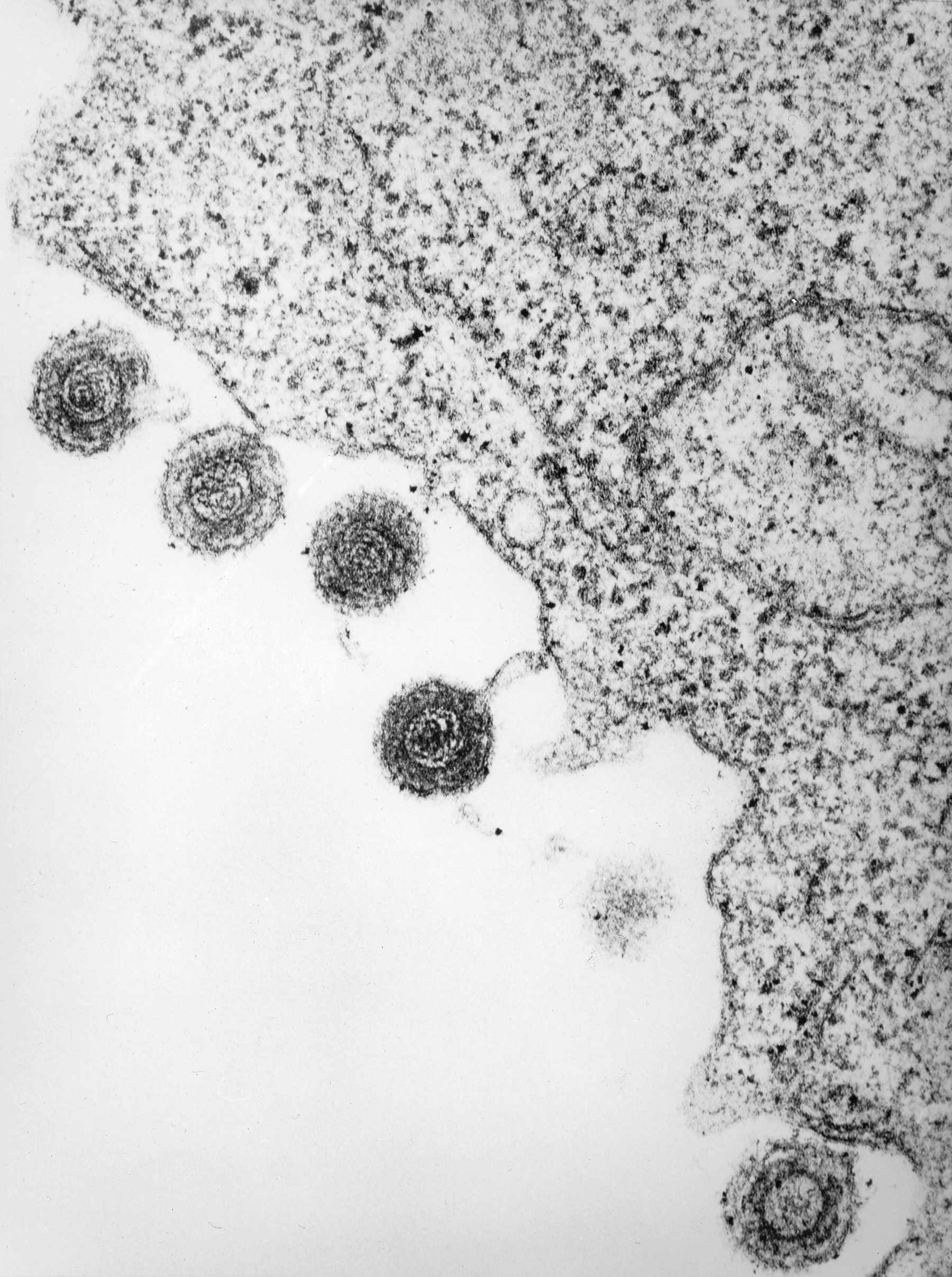
This electron micrograph shows herpes virus type 6 (HHV-6) maturing and being released from a white blood cell. HHV-6 is a double-stranded DNA virus.

Credit

Public domain/ Laboratory of Tumor Cell Biology, National Cancer Institute/ Bernard Kramarsky, photographer/ 1986

Source

http://visualsonline.cancer.gov/details.cfm?imageid=2256

Image wall, Microbe SEMS

Human herpes virus type 6

January 9, 2013 jbrehm2
Description

This electron micrograph shows human herpes virus type 6 (HHV-6), which is a double-stranded DNA virus.

Credit

Public domain/ Laboratory of Tumor Cell Biology, National Cancer Institute/ Bernard Kramarsky, photographer/ 1986

Source

http://visualsonline.cancer.gov/details.cfm?imageid=2257

Image wall, Microbe SEMS

Electron micrograph of herpes simplex virus

January 9, 2013 jbrehm2
Description

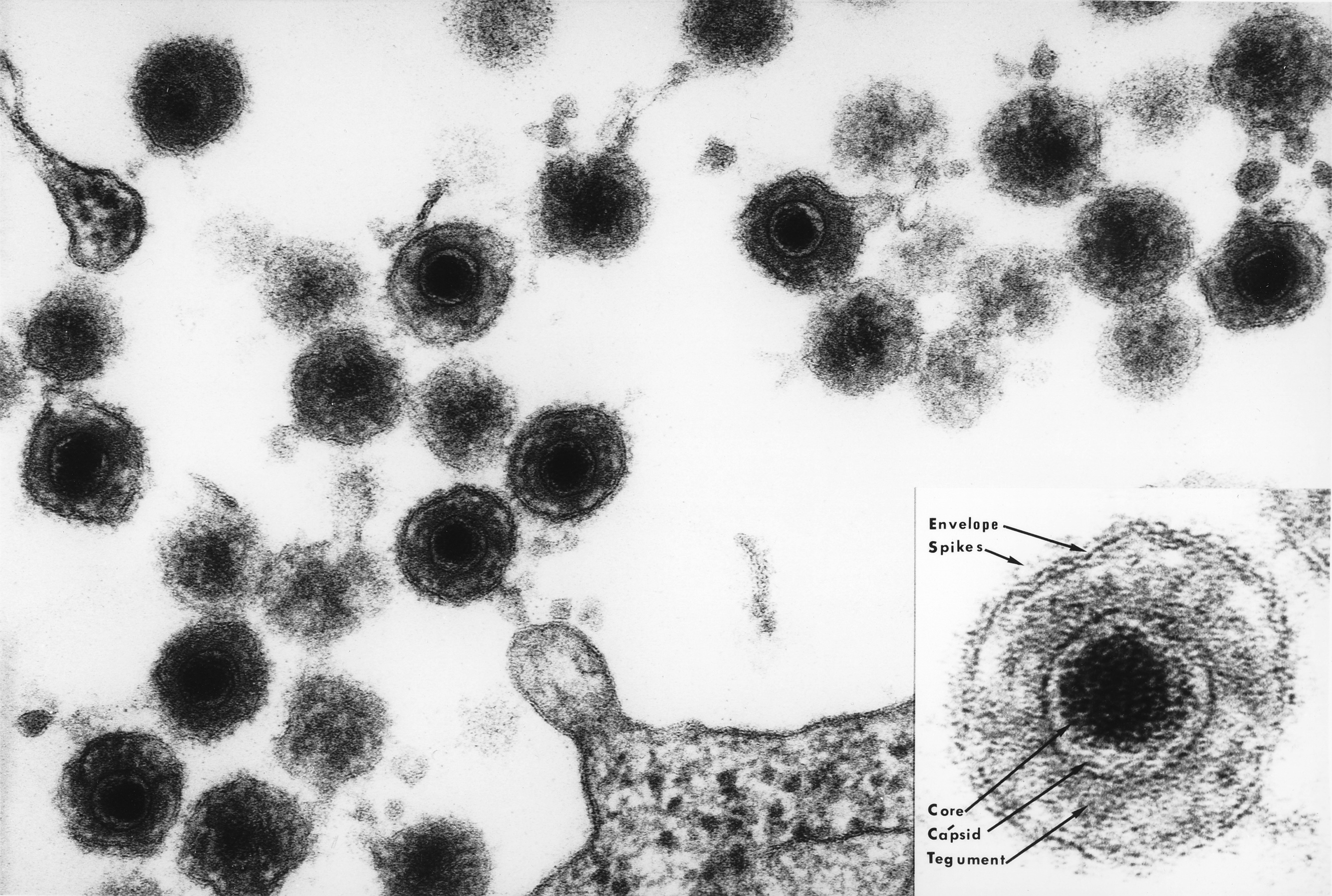
This electron micrograph shows numerous virions of herpes simplex virus.

Credit

Public domain/ Centers for Disease Control and Prevention/ Dr. Fred Murphy and Sylvia Whitfield, content providers/ 1975

Source

http://phil.cdc.gov/phil/imageidsearch.asp (Image ID # 10230)

Image wall, Microbe SEMS

Electron micrographs of herpes viruses

January 9, 2013 jbrehm2
Description

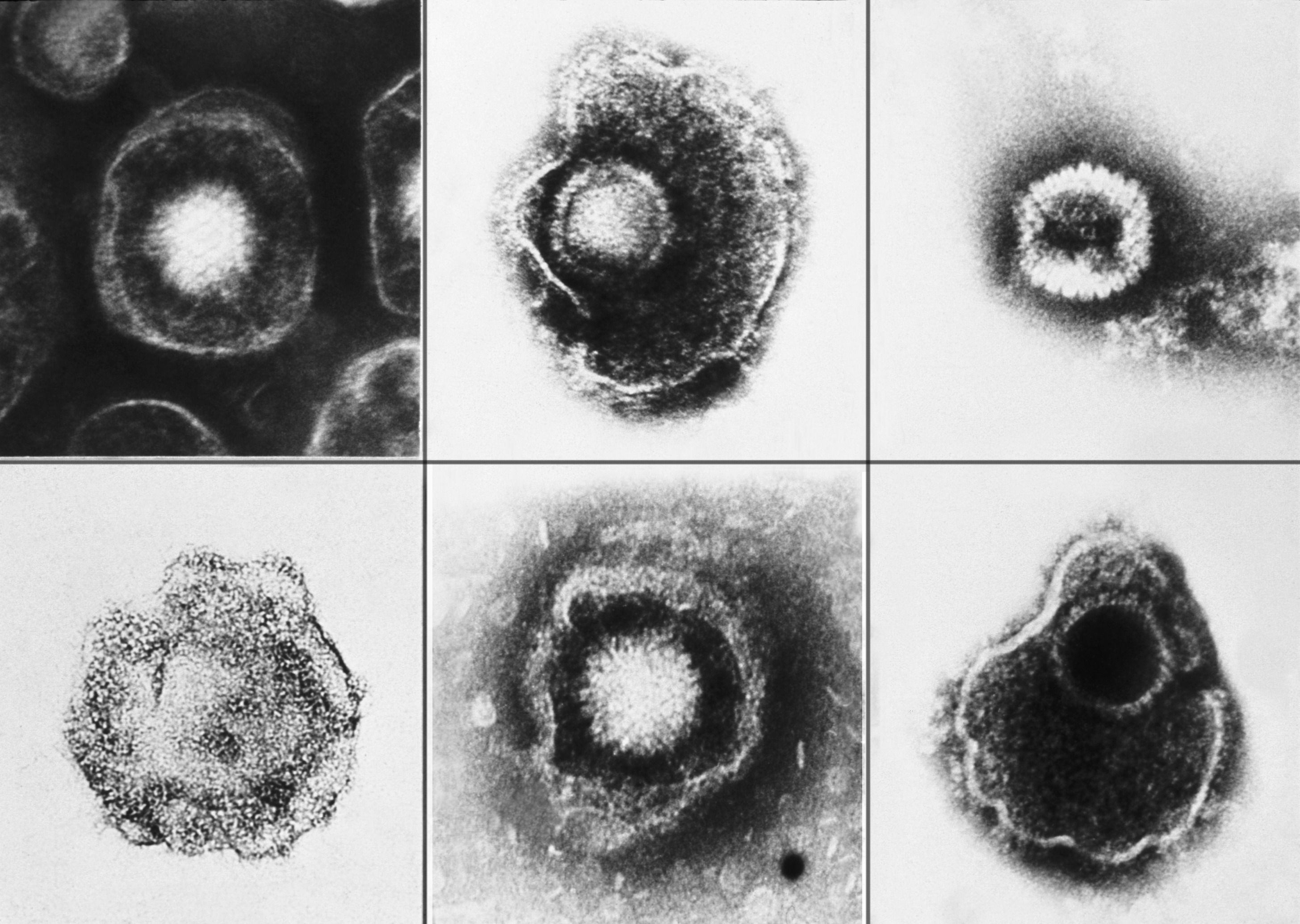
These electron micrograph images show herpes viruses, including varicella-zoster virus (chickenpox virus), and herpes simplex virus types 1 and 2 (HSV-1, HSV-2).

Credit

Public domain/ Centers for Disease Control and Prevention/ E. L. Palmer, content provider/ 1981

Source

http://phil.cdc.gov/phil/imageidsearch.asp (Image ID # 2171)

Image wall, Microbe SEMS

Electron micrograph of Epstein-Barr virus

January 9, 2013 jbrehm2
Description

This electron micrograph image shows Epstein-Barr viruses.

Credit

Public domain-freely reused/ Laboratory of Tumor Cell Biology/ National Cancer Institute/ Linda Bartlett, photographer/ 1980

Source

http://visualsonline.cancer.gov/details.cfm?imageid=2105

Image wall, Microbe SEMS

Epstein-Barr viruses

January 9, 2013 jbrehm2
Description

This electron micrograph shows numerous Epstein-Barr virus particles. This virus contains a double-stranded DNA linear genome.

Credit

Public domain/ Centers for Disease Control and Prevention/ Dr. Fred Murphy, content provider/ 1975

Source

http://phil.cdc.gov/phil/imageidsearch.asp (Image ID # 10234)

Image wall, Microbe SEMS

Herpes simplex virions inside a cell

January 9, 2013 jbrehm2
Description

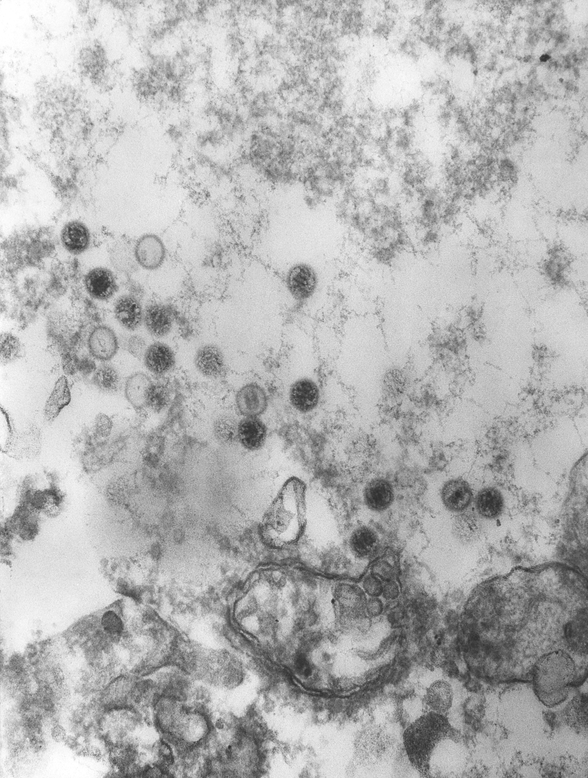
This electron micrograph shows numerous herpes simplex virions located inside a cell nucleus.

Credit

Public domain/ Centers for Disease Control and Prevention/ Dr. Fred Murphy and Sylvia Whitfield, content providers/ 1975

Source

http://phil.cdc.gov/phil/imageidsearch.asp (Image ID # 10261)

Image wall, Microbe SEMS

Electron micrograph of herpes simplex virions

January 9, 2013 jbrehm2
Description

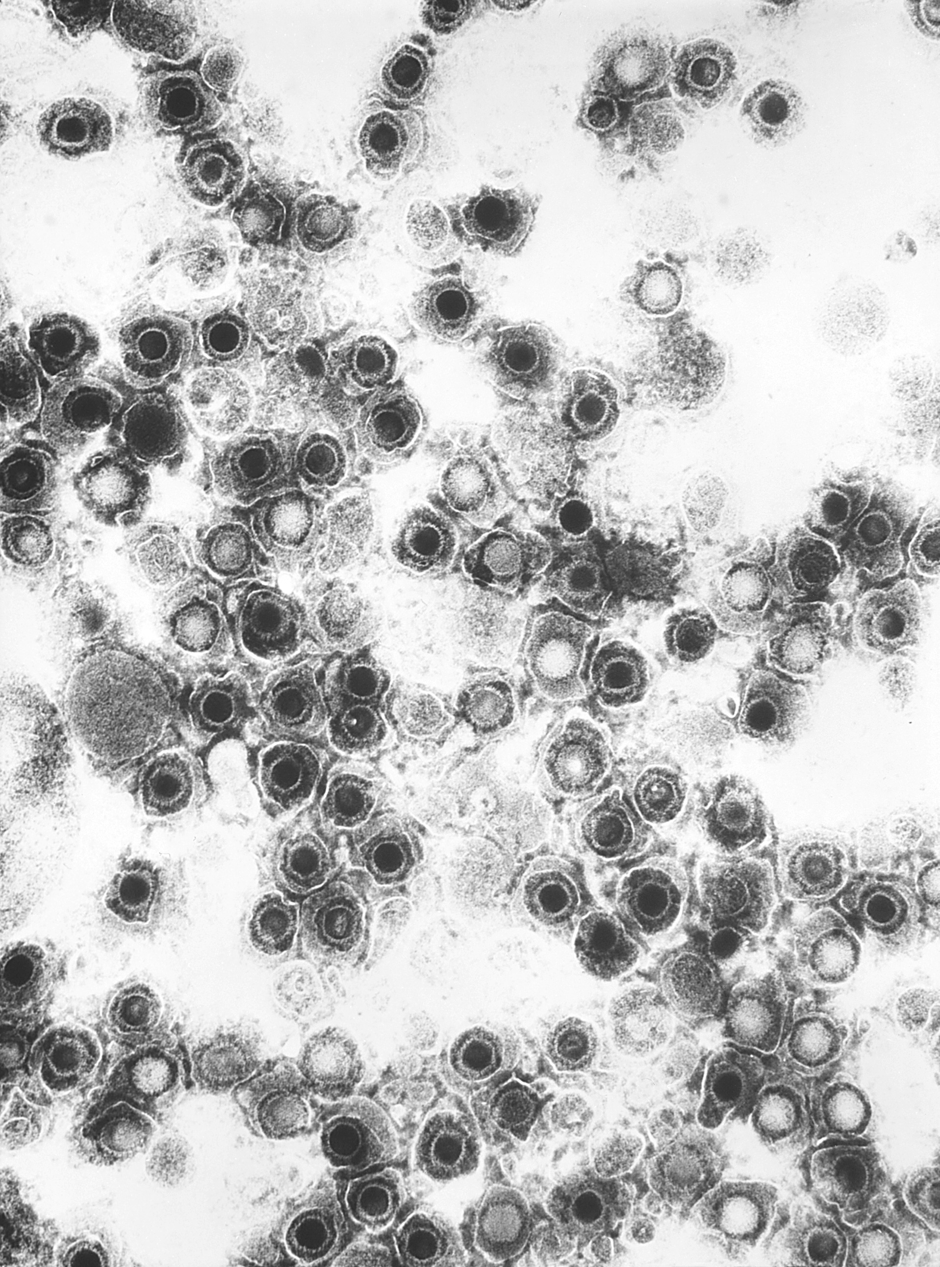
This electron micrograph shows numerous herpes simplex virions.

Credit

Public domain/ Centers for Disease Control and Prevention/ Dr. Fred Murphy and Sylvia Whitfield, content providers/ 1975

Source

http://phil.cdc.gov/phil/imageidsearch.asp (Image ID # 10232)

Posts navigation

← Previous 1 2 3 … 18 Next →

Recent Posts

  • Tribal C’rona Comix Page 5
  • Tribal C’rona Comix Page 4
  • Tribal C’rona Comix Page 3
  • Tribal C’rona Comix Page 2
  • Tribal C’rona Comix Page 1

Recent Comments

    Archives

    • December 2020
    • November 2020
    • October 2020
    • September 2020
    • August 2020
    • July 2020
    • June 2020
    • February 2015
    • December 2014
    • January 2013

    Categories

    • Apps and Activities
    • Bacterial Viruses
    • Covid-19
    • Crona Comix II
    • Curricula
    • Dengue Virus
    • Ebola Virus
    • Epidemic Simulations
    • Foot and Mouth Disease Virus
    • General
    • Hantavirus
    • Hepatitis B Virus
    • Historical Images
    • HIV
    • HPV
    • Image wall
    • Influenza Virus
    • Inside Viruses
    • Microbe Illustrations
    • Microbe Scientists
    • Microbe SEMS
    • Polio Virus
    • Rabies Virus
    • SARS Virus
    • Tribal Crona Comix
    • Uncategorized
    • West Nile Virus

    Meta

    • Log in
    • Entries feed
    • Comments feed
    • WordPress.org