Marine viruses, shown in purple, surround potential microbes. The odds are stacked against the microbes when there are so many viruses. Willie Wilson, Bigelow Laboratory for Ocean Sciences.

Phages are viruses that infect bacteria. They have a characteristic shape. Cyanophage photo by Willie Wilson, Bigelow Laboratory for Ocean Sciences.
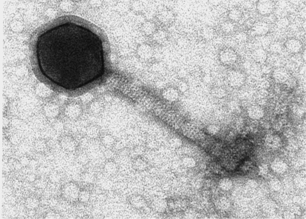
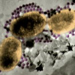
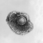
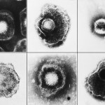
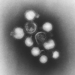
This electron micrograph shows Coxsackie B4 virus particles.
Public domain/ Centers for Disease Control and Prevention/ 1981
http://phil.cdc.gov/phil/imageidsearch.asp (Image ID # 5630)
Low res High res
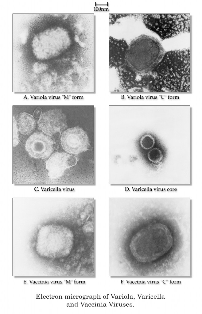
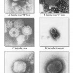
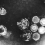
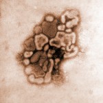
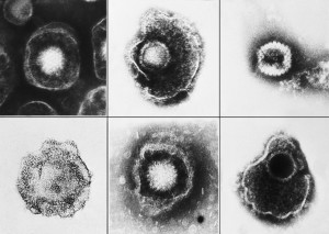
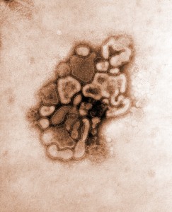
This electron micrograph shows variola, varicella, and vaccinia virions.
Public domain/ Centers for Disease Control and Prevention/ Dr. James Nakano, content provider/ 1975
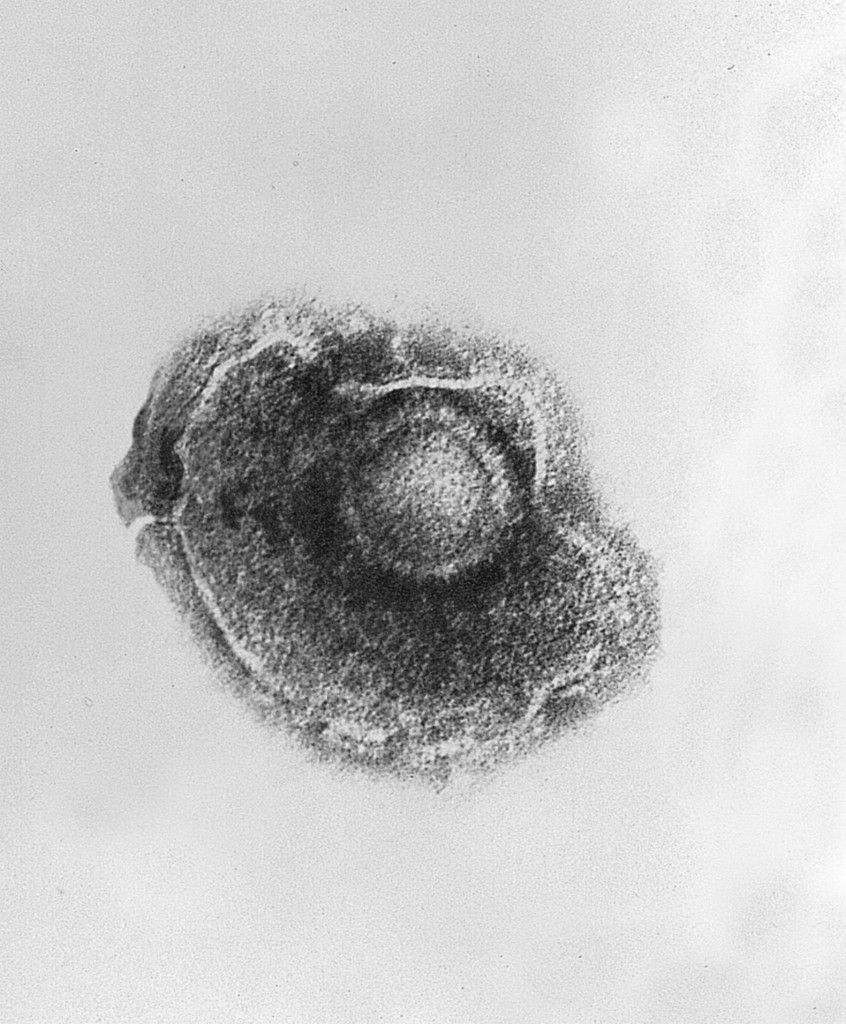
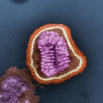
This electron micrograph shows the varicella (chickenpox) virus.
Public domain/ Centers for Disease Control and Prevention/ Dr. Erskine Palmer and B.G. Partin, content providers/ 1982
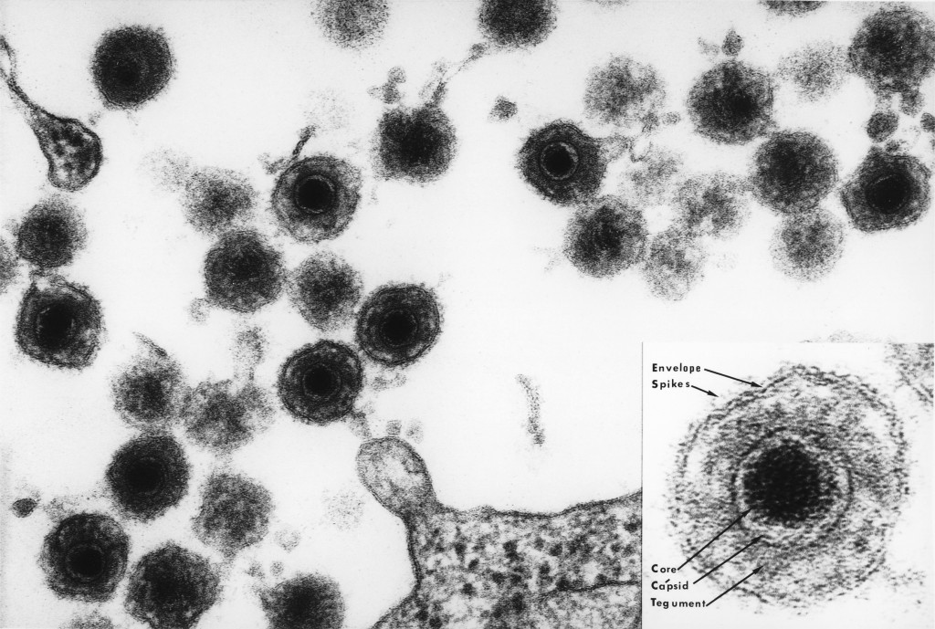
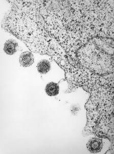
This electron micrograph shows herpes virus type 6 (HHV-6) maturing and being released from a white blood cell. HHV-6 is a double-stranded DNA virus.
Public domain/ Laboratory of Tumor Cell Biology, National Cancer Institute/ Bernard Kramarsky, photographer/ 1986
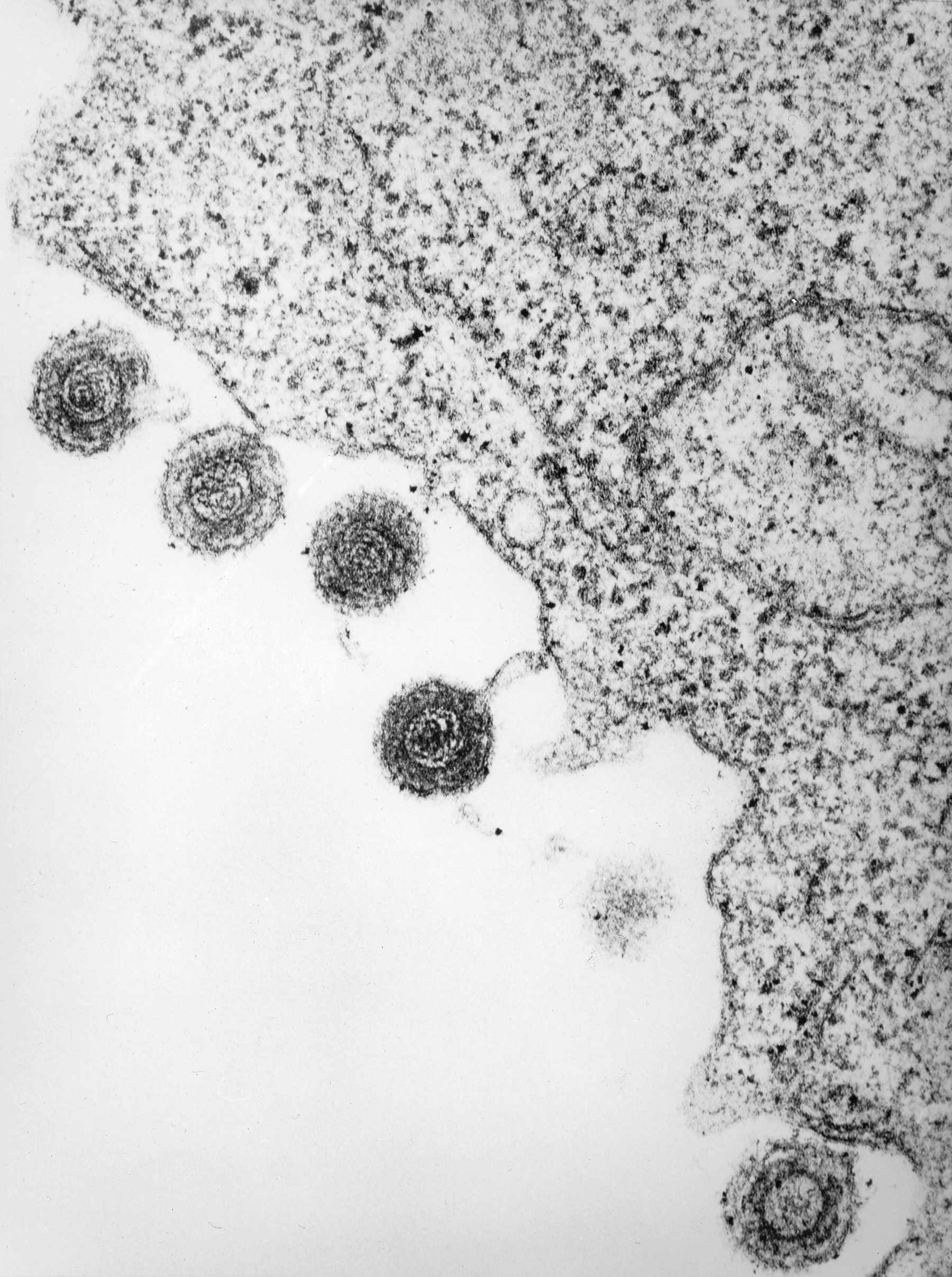
This electron micrograph shows human herpes virus type 6 (HHV-6), which is a double-stranded DNA virus.
Public domain/ Laboratory of Tumor Cell Biology, National Cancer Institute/ Bernard Kramarsky, photographer/ 1986
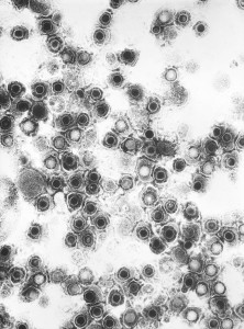
This electron micrograph shows numerous virions of herpes simplex virus.
Public domain/ Centers for Disease Control and Prevention/ Dr. Fred Murphy and Sylvia Whitfield, content providers/ 1975

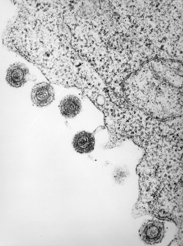
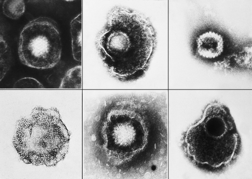
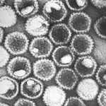
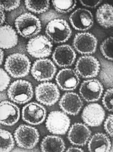
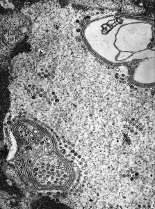
These electron micrograph images show herpes viruses, including varicella-zoster virus (chickenpox virus), and herpes simplex virus types 1 and 2 (HSV-1, HSV-2).
Public domain/ Centers for Disease Control and Prevention/ E. L. Palmer, content provider/ 1981
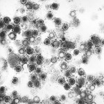
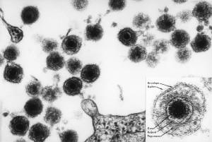
This electron micrograph image shows Epstein-Barr viruses.
Public domain-freely reused/ Laboratory of Tumor Cell Biology/ National Cancer Institute/ Linda Bartlett, photographer/ 1980

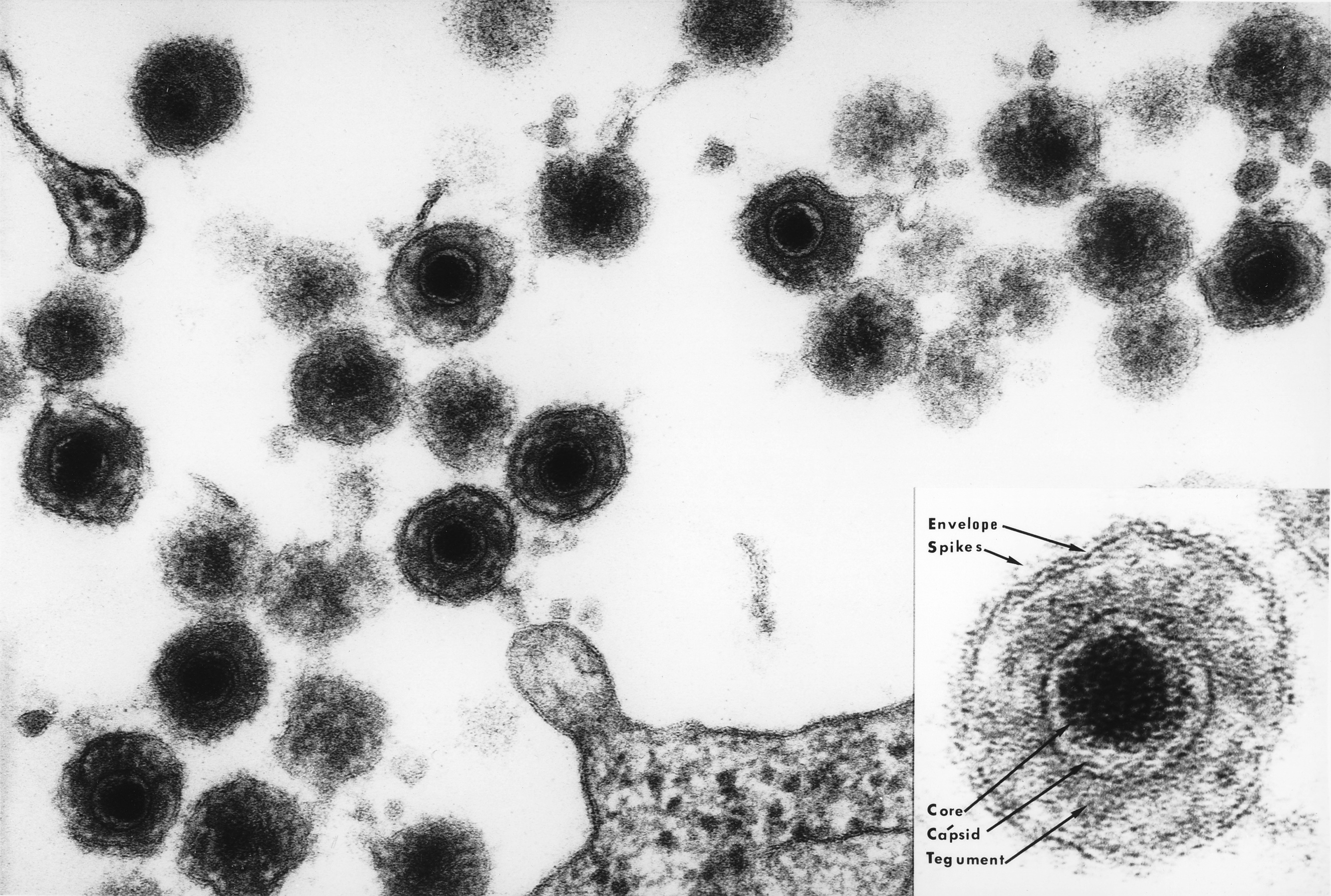
This electron micrograph shows numerous Epstein-Barr virus particles. This virus contains a double-stranded DNA linear genome.
Public domain/ Centers for Disease Control and Prevention/ Dr. Fred Murphy, content provider/ 1975

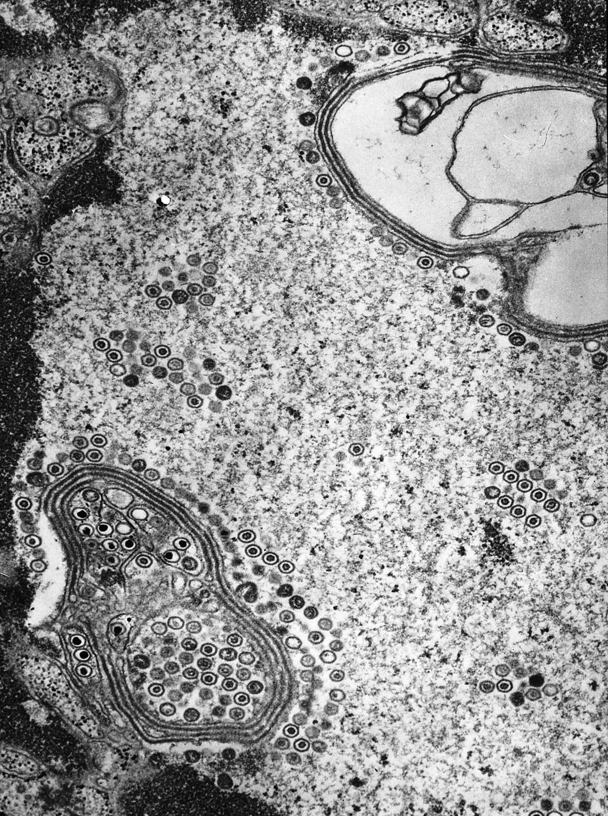
This electron micrograph shows numerous herpes simplex virions located inside a cell nucleus.
Public domain/ Centers for Disease Control and Prevention/ Dr. Fred Murphy and Sylvia Whitfield, content providers/ 1975

This electron micrograph shows numerous herpes simplex virions.
Public domain/ Centers for Disease Control and Prevention/ Dr. Fred Murphy and Sylvia Whitfield, content providers/ 1975
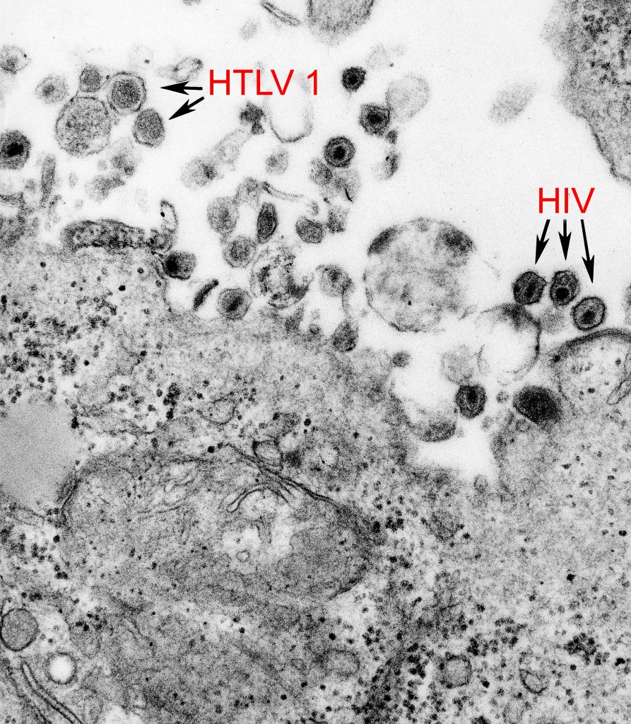
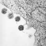
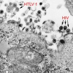
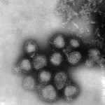
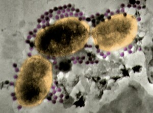
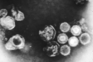
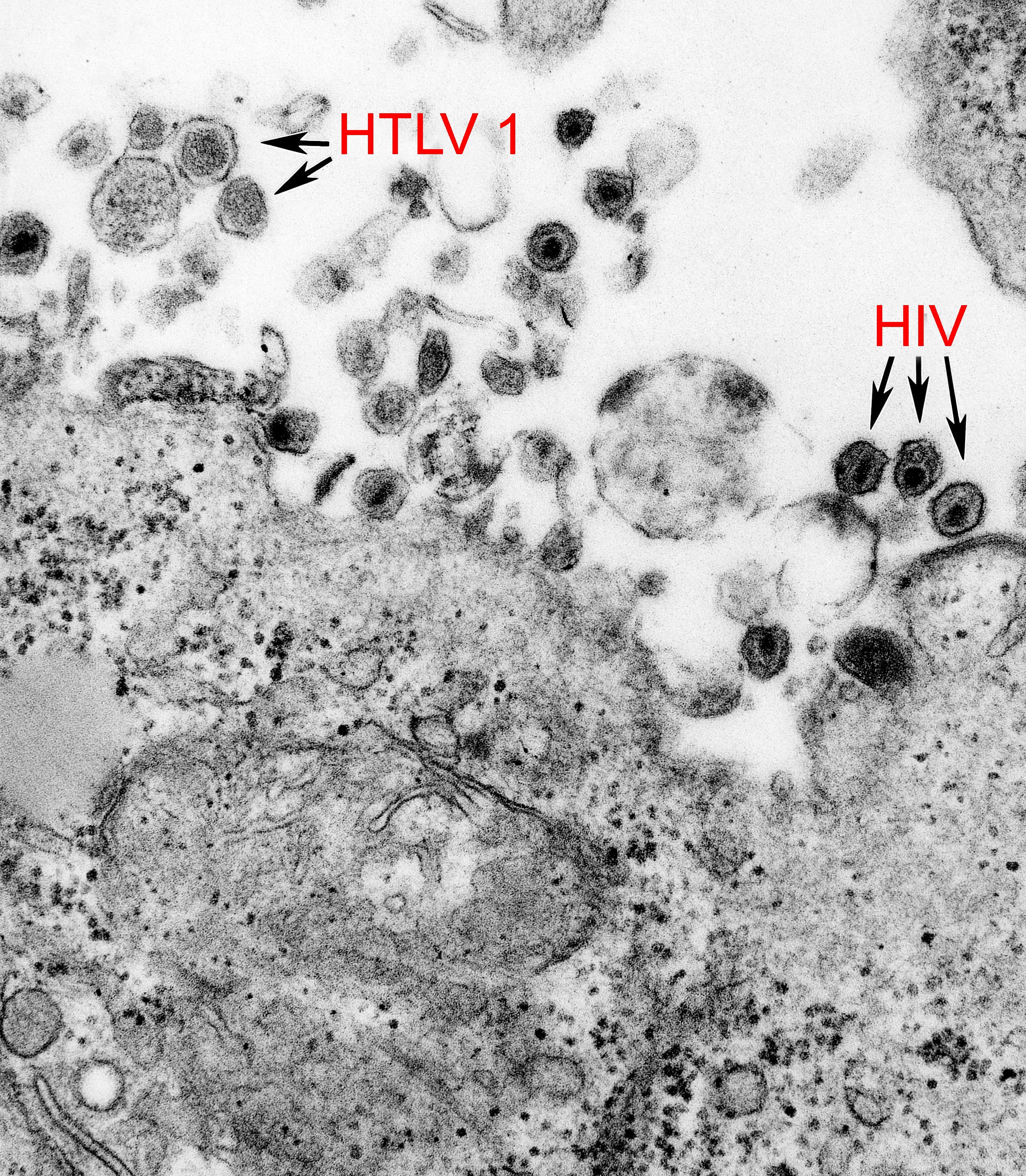
This electron micrograph shows both the human T-cell leukemia type-1 virus (HTLV-1) and the human immunodeficiency virus (HIV).
Public domain/ Centers for Disease Control and Prevention/ Cynthia Goldsmith, content provider/ Unknown date
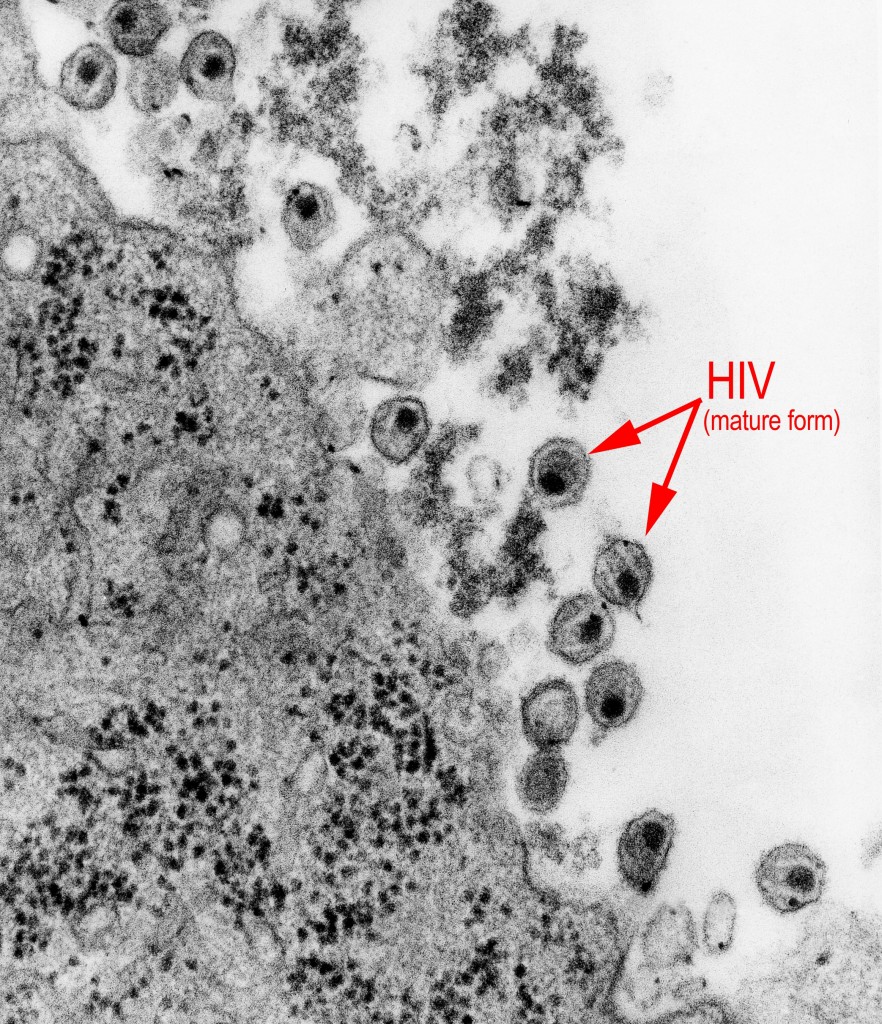
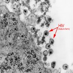
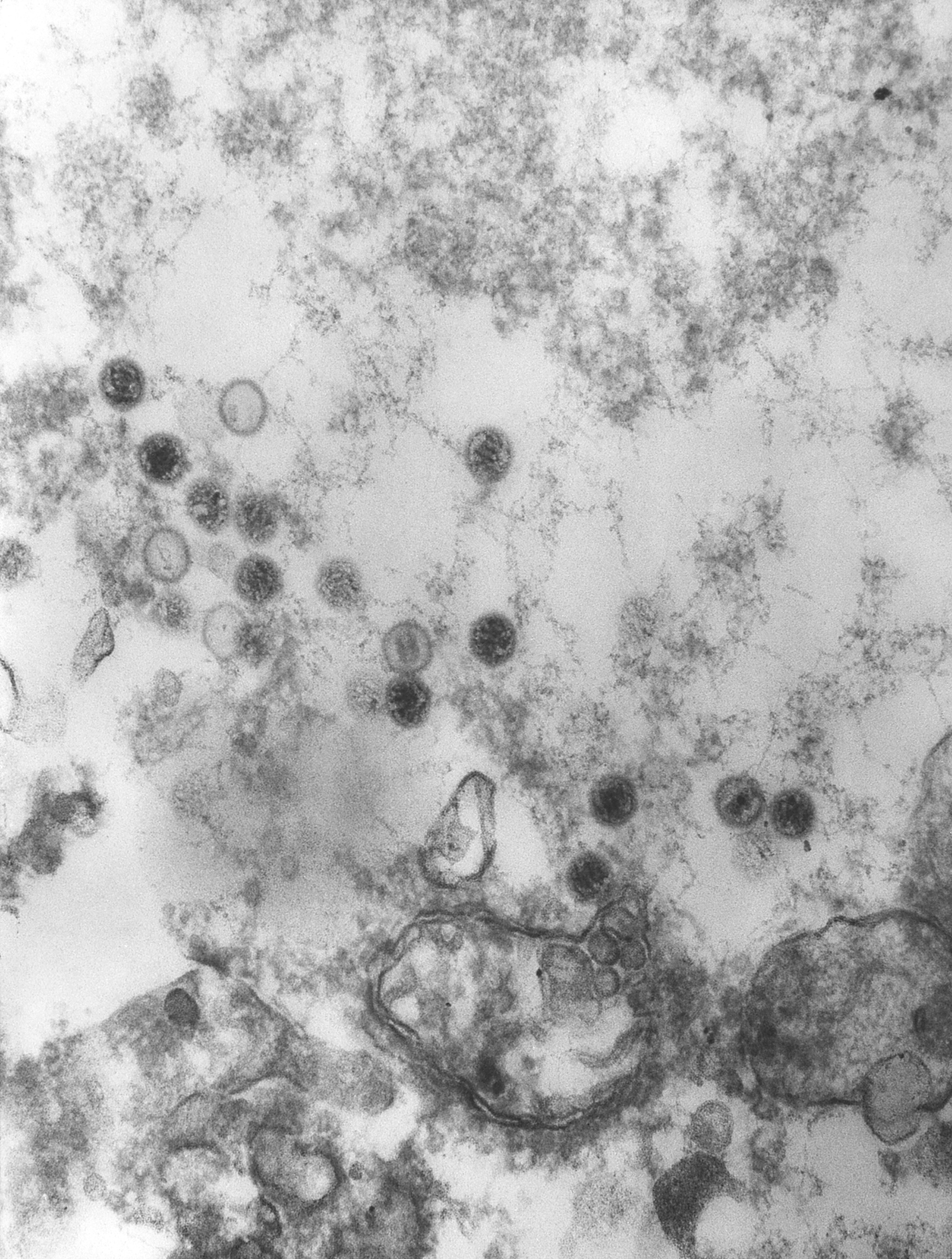
This electron micrograph shows mature forms of the human immunodeficiency virus (HIV).
Public domain/ Centers for Disease Control and Prevention/1983
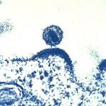
This electron photomicrograph shows HIV budding out of a human immune cell, which the virus infects and uses to make copies.
Public domain/ National Institute of Allergy and Infectious Diseases, National Institutes of Health/ courtesy of Dr. Tom Folks/ 2008

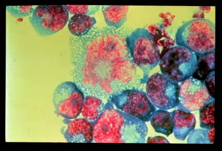
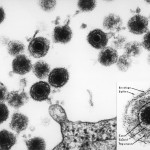
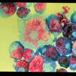
This electron photomicrograph shows T cells infected with HIV.
Public domain / National Institute of Allergy and Infectious Diseases, National Institutes of Health / Department of Health and Human Services, courtesy of Dr. Tom Folks / 2008
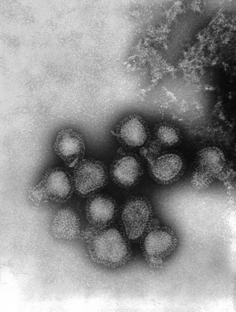
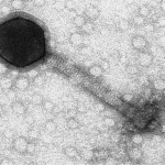
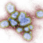
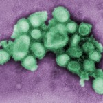
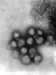
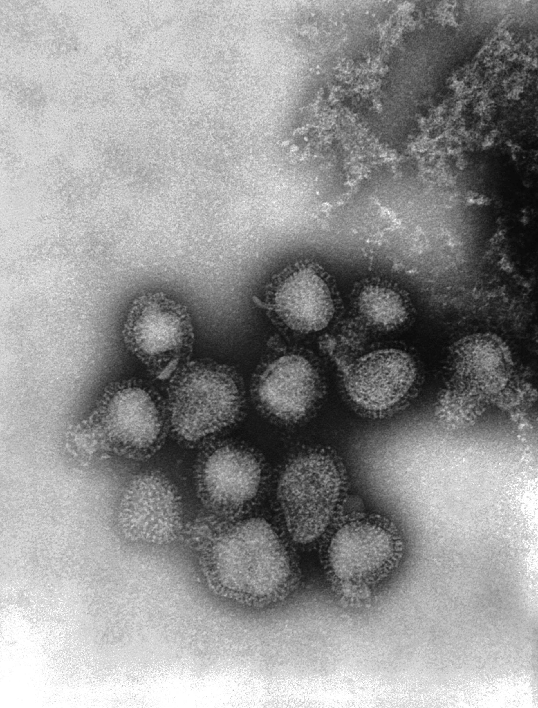
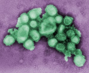
Electron micrograph shows a Hong Kong H3N2 subtype of the influenza A virus.
Public domain/ Centers for Disease Control and Prevention/ Dr. Fred Murphy, content provider/ 1975
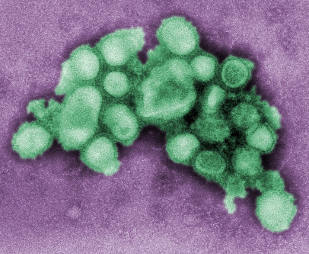
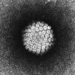
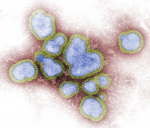
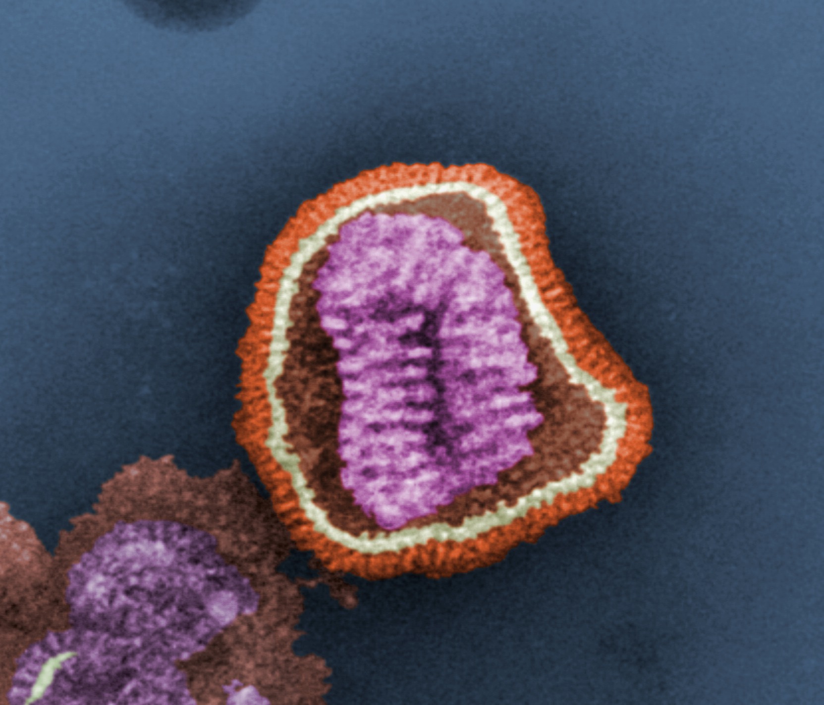
This electron micrograph shows a number of influenza virions. Notice the neuraminidase (N) and hemagglutinin (H) spikes sticking out from the protein coat, or capsid.
Public domain/ Centers for Disease Control and Prevention/ Dr. John Hierholzer, content provider/ 1974

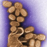
This electron micrograph shows an H1N1 influenza virus that causes disease in pigs. It is magnified 37,800 times. Several virus particles are developing while being grown in chicken eggs.
Public domain/ Centers for Disease Control and Prevention/ Dr. E. Palmer and R.E. Bates, content providers/ 1976

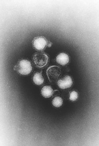
This electron micrograph shows virus particles, or virions, from Type A influenza virus.
Public domain/ Centers for Disease Control and Prevention/ F.A. Murphy, content provider/ 1976

This electron micrograph shows several virus particles, or virions, of a re-created 1918 influenza virus growing in kidney cells of a dog.
Public domain/ Centers for Disease Control and Prevention/ Dr. Terrence Tumpey, content provider/ Cynthia Goldsmith, photographer/ 2005
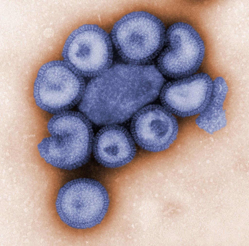
Electron micrograph shows a number of influenza virus particles, or virions.
Public domain/ Centers for Disease Control and Prevention/ Dr. F. A. Murphy, content provider/ 1973
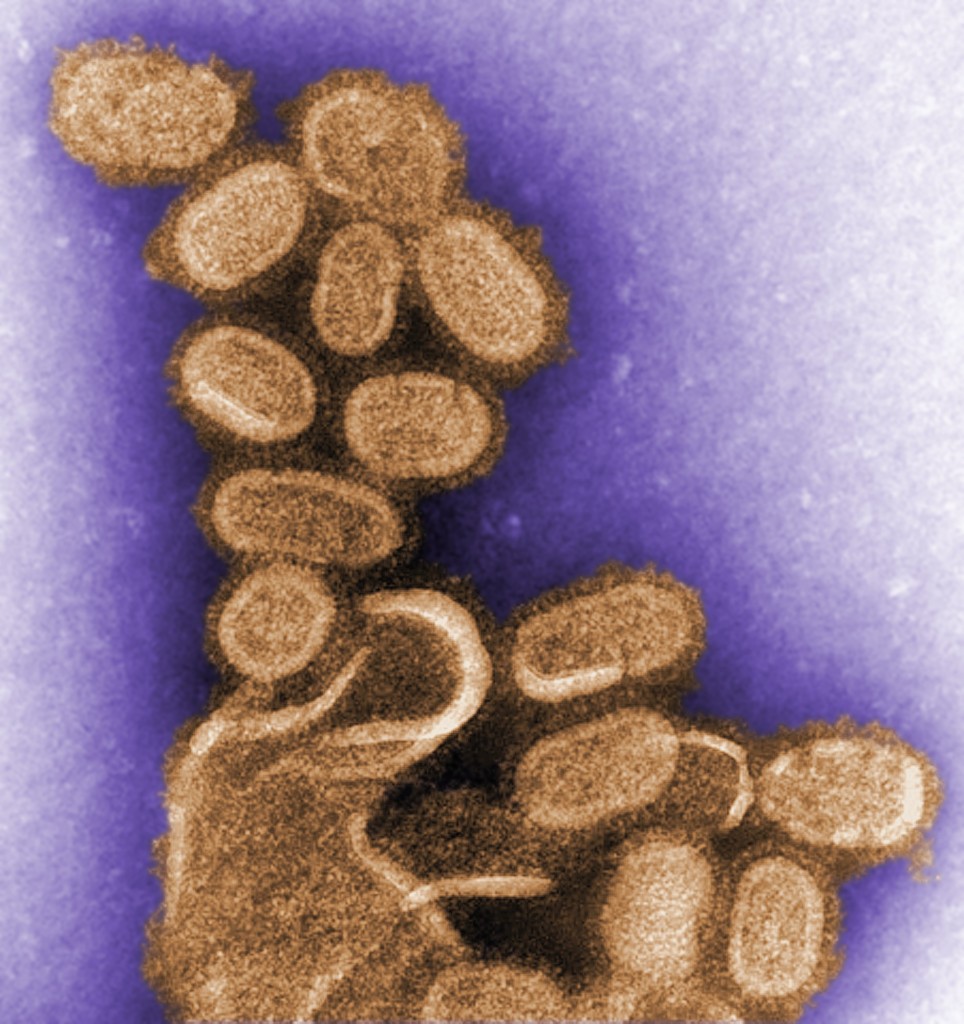
This electron micrograph shows several particles, or virions, of an influenza virus that causes the disease in pigs.
Public domain/ Centers for Disease Control and Prevention/ C. S. Goldsmith and A. Balish, content providers/ 2009
This electron micrograph shows an influenza virus particle, or virion. Influenza is a single-stranded RNA virus.
Public domain/ Centers for Disease Control and Prevention/ E. L. Palmer and M. Larkin, content providers/ F. Murphy, photographer/ 1981

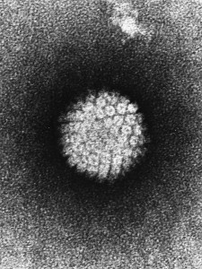
This is an electron photomicrograph of human papillomavirus (HPV) occurring in human warts.
Public domain/ Laboratory of Tumor Virus Biology, National Cancer Institute/ 1986

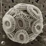
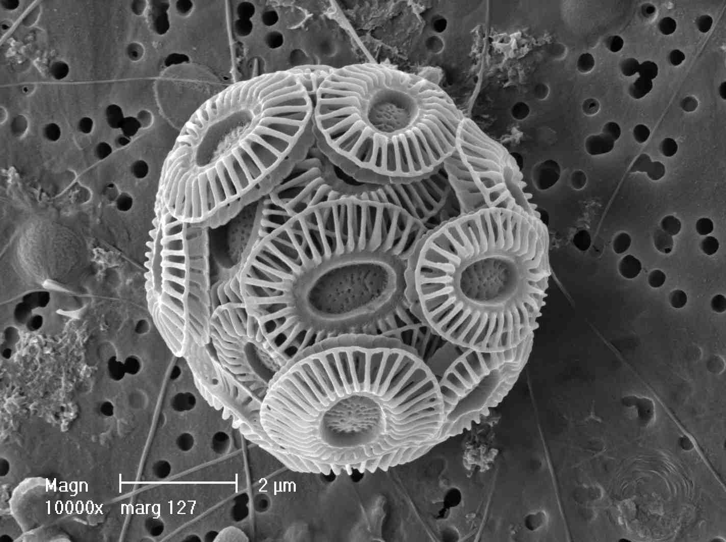
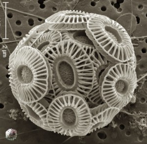
This electron micrograph of the algae, Emiliania huxleyi, shows the size comparison with the much smaller Emiliania huxleyi virus (EhV) at the lower left corner.
Permission granted for educational use/ The Natural History Museum, London (Dr. Jeremy Young) and University of Nebraska-Lincoln (Angie Fox) / 2009
http://www.noc.soton.ac.uk/soes/staff/tt/eh/pics/cocco9.jpg
Low res High res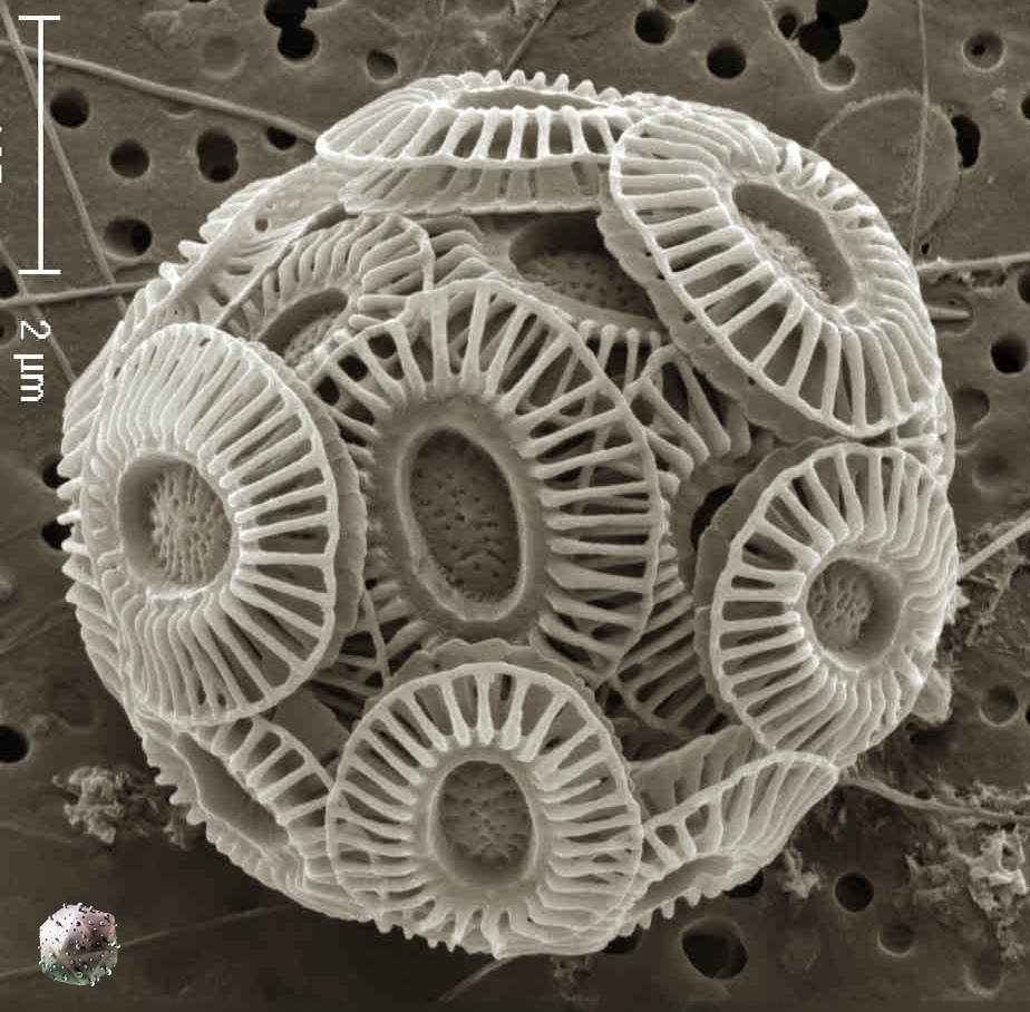


